

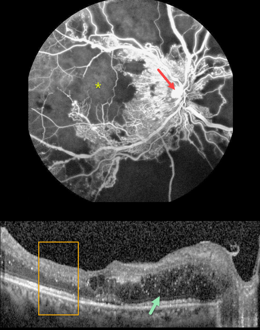
This 27YO female with a history of uncontrolled type 1 diabetes presented with bilateral vision loss. Vision was 20/40 OD and 20/200 OS.
Fluorescein angiography shows severe capillary loss that extends through the central macula. The remaining vessels are telangiectatic, especially bordering the areas of frank loss, and an area of leaking disc neovascularization is noted.
OCT scanning shows mostly nasal edema of the outer nuclear layer with numerous suspended hyperreflective particles (hyperreflective foci, HRF), along with more central cysts and trace subretinal fluid. The inner retinal layers have lost some of their normal hyper- and hypo-reflective structures (disorganization of the retinal inner layers, DRIL). Similar findings were noted in her left eye (images not shown).
Monthly anti-VEGF therapy was recommended although the patient was subsequently lost to follow-up.
Learning Points:
Anti-VEGF therapy has become the standard treatment for central-involved diabetic macular edema (CI-DME), with the vast majority of eyes showing improvements in macular thickness and vision.
Although we expect our patient’s edema to improve if she returns for treatment, we are more guarded regarding her final acuity since the capillary loss is permanent. Eyes with CI-DME and baseline central DRIL have a worse visual prognosis (Sun et al, JAMA Ophthalmol 2014;123:1309-1316).
HRF are small dot-like retinal opacities found in some eyes with diabetic macular edema. Although their pathogenesis is debated, they likely represent extravasated lipoproteins that can later become clinically evident lipid exudates (Ganne et al, Indian J Ophthalmol 2021;69:3208-3217).
Although HRF numbers decrease with treatment, it is unclear whether they serve as a biomarker for predicting visual outcome (Huang et al, Ophthalmology Retina 2022;6:814-827).



