

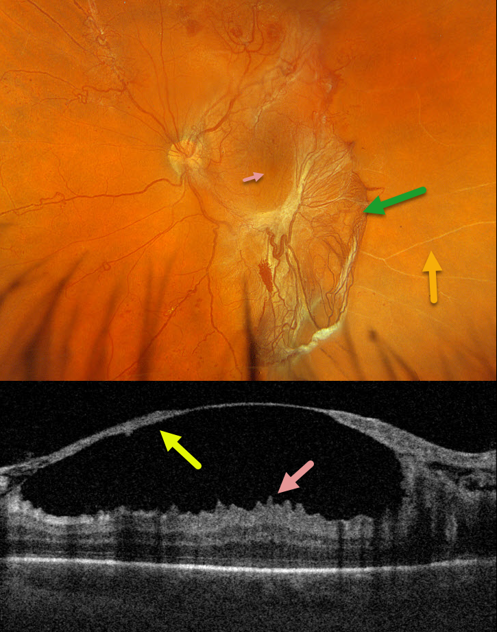
This 42YO male with type 2 diabetes presented with 1 week of vision loss in his left eye. Vision was 20/40 OD and 20/100 OS.
Optos color RGB imaging shows a large patch of variably fibrosed retinal neovascularization extending from the temporal macula into the inferior midperiphery. Vertical macular striae are drawn towards the neovascularization inferiorly. Scattered neovascularization is seen elsewhere, along with nonperfused large retinal vessels temporally.
Triton swept-source OCT through the macular center shows a thickened detached posterior hyaloid that extends from the optic nerve into the temporal macula. The macular striae noted on the Optos image correspond to inner macular folds on OCT. Vitrectomy surgery was scheduled.



