

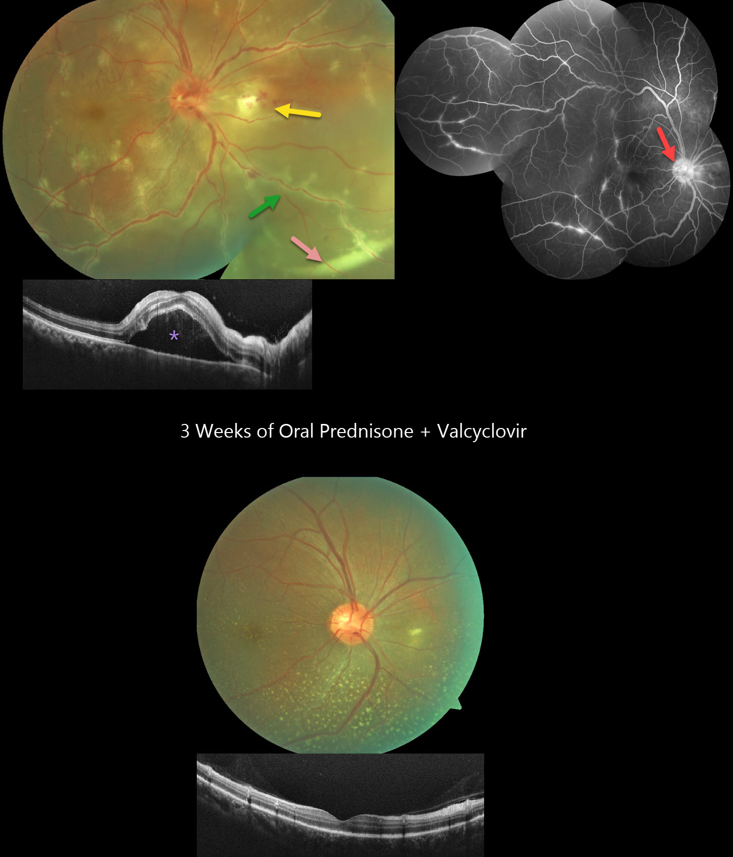
This healthy 36YO male presented with 3 days of floaters and decreased vision in his right eye. Vision was 20/60 OD and 20/20 in his healthy OS. The right eye contained mild anterior chamber cells and moderate vitreous cells.
Fundus photography shows a swollen nerve, a frosted branch pattern of vasculitis involving the retinal veins, an area of retinitis just nasal to the nerve, and a subretinal hypopyon in the inferonasal midperiphery. Some multifocal areas of outer retinal inflammation are noted, along with some retinal hemorrhages. OCT scanning shows variable hyperreflective foveal subretinal fluid. Fluorescein angiography shows variable venous staining and leakage, including from the disc and in nasal retinitis.
The workup included elevated ESR and CRP levels, along with an increased IgG titer for Varicella zoster virus. Mantoux testing, syphilis, Bartonella, typhus, HIV, and chest X-ray were all negative. Although very atypical for viral uveitis, we started oral prednisone and valaciclovir. After one week, the hypopyon resolved, with decreased vasculitis and new scattered flecks of subretinal lipid throughout the posterior pole. By three weeks, vision improved to 20/20 with continued funduscopic improvement.
Learning Points:
Epidemic (post-fever) retinitis is an infectious or para-infectious entity that develops in immunocompetent individuals several weeks following a bacterial, viral, or protozoal infection. Causes include Dengue, Rickettsiosis, West Nile, Leptospirosis, Chikungunya, Typhoid, and Ebola. Patients present with sudden, painless vision loss.
Fundus findings include unilateral or bilateral unifocal or multifocal patches of retinitis with possible nerve involvement, serous macular detachment, and vasculitis or frosted branch angiitis. There is usually a favorable response to steroids and treating the underlying infectious cause with appropriate systemic therapy. See Mahendradas et al (Indian J Ophthalmol 2020;68:1775-1786) for an excellent post-fever retinitis review.
Frosted branch angiitis is a retinal vasculitis that gets its name from the involved vessels resembling branches of a tree that are “frosted” with snow (Kleiner et al, AJO 1988;106:27-34). Although these findings may be isolated, they are often considered a phenotype found in many inflammatory conditions, most commonly CMV retinitis. The inflammation is usually highly responsive to steroids, and the visual prognosis is generally good, although some may develop secondary neovascularization in the anterior and posterior segments due to widespread ischemia.
Subretinal hypopyon is characterized by yellowish inflammatory material settling inferiorly in the subretinal space. It has been documented in infectious (bacterial and fungal endophthalmitis, acute retinal necrosis, tuberculosis, syphilis), inflammatory (sympathetic ophthalmia), and



