

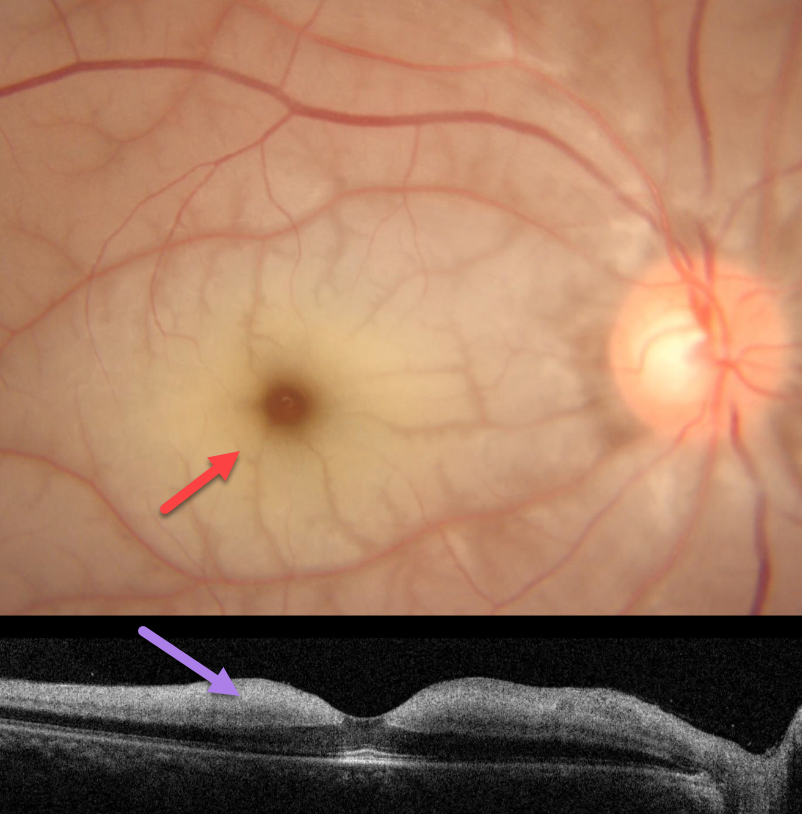
This 41YO male presented with a 2-day history of acute vision loss in his right eye. Vision was counting fingers OD and 20/20 in his normal OS.
Fundus photography shows an acute central retinal artery occlusion (CRAO) with a pale, opaque inner retina and a cherry-red spot. The retina immediately adjacent to the radiating perifoveal arterioles is relatively transparent, creating a fern-like appearance. OCT shows a slightly thickened and markedly hyperreflective inner retina. The patient was immediately sent to the nearest stroke center.
Learning Points:
Acute CRAO often exists along a spectrum from complete occlusion with a stagnant blood column to a more partial blockage with variable patchy ischemia. Our patient still has enough active flow to supply the neurosensory retina immediately adjacent to the smaller arterioles, but not enough to supply the more distal retinal tissue.
About 90% of CRAO are embolic, with the remainder most commonly due to giant cell arteritis. The lack of a visible embolus, as in our patient, does not preclude an embolic cause since the causative embolus can either be posterior to the visible disc or have already traveled further downstream.



