

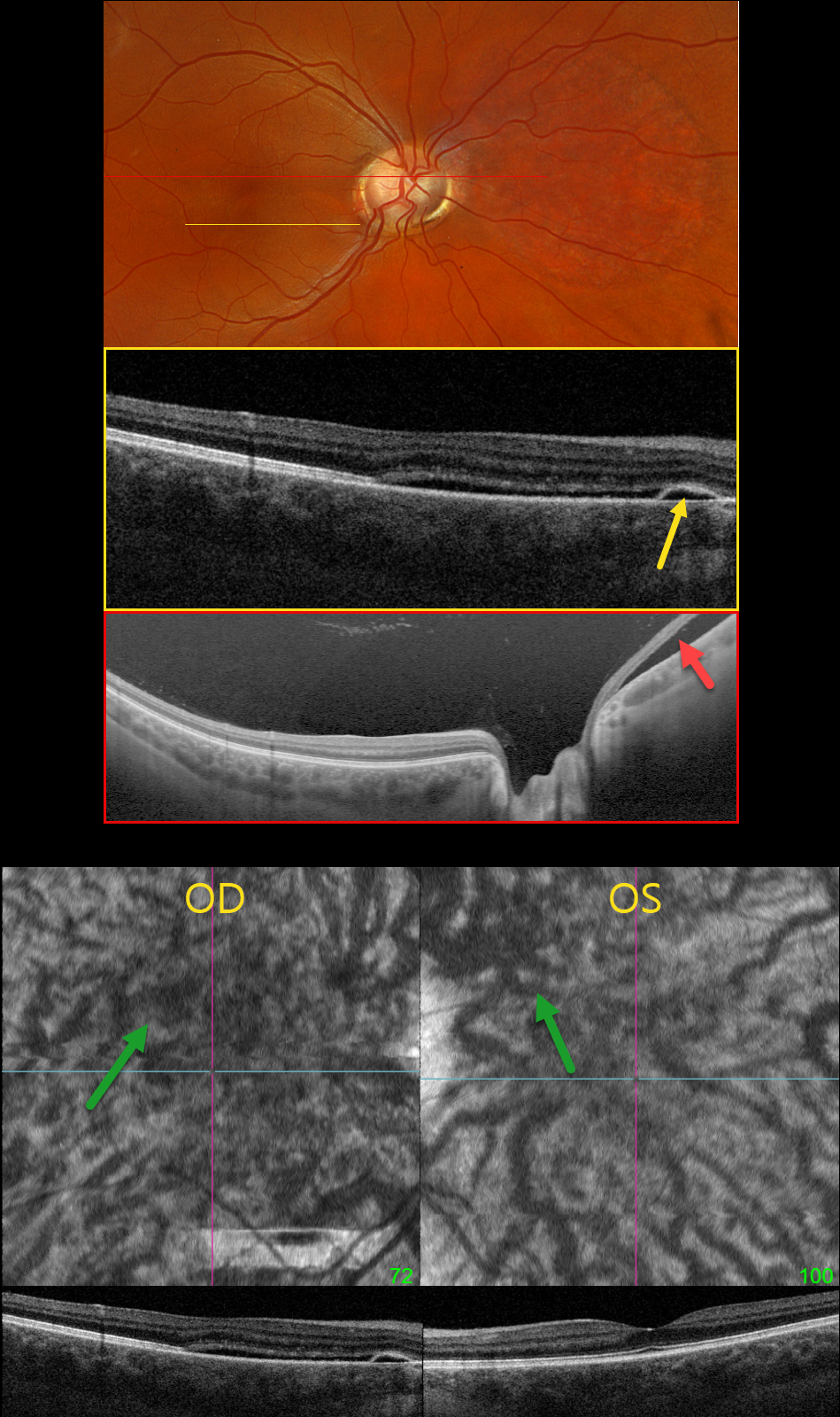
This healthy pregnant 32YO female presented with recent mild central vision loss in her right eye. Vision was 20/40 OD and 20/30 OS.
Optos color RGB imaging shows an anomalous right optic disc with an optic pit both nasally and temporally. Triton swept-source OCT shows both pits and subretinal fluid extending from the nerve nasally. OCT through the macula shows shallow nasal subretinal fluid extending into the macular center, along with a tiny nasal RPE detachment. This fluid does not communicate with the nerve or temporal pit. Optos Silverstone swept-source OCT through the posterior pole shows the subretinal fluid nasal to the nerve. Macular en face and B-scans show a thickened choroid with anomalous, thickened, and tortuous vessels.
Our patient’s symptoms are from idiopathic central serous retinopathy (ICSC), most likely related to her pregnancy. The macular fluid does not communicate with the temporal disc pit, and her macular OCT findings are classic for central serous. Fluorescein angiography was not performed due to her pregnancy. Although she has optic pit ‘maculopathy,’ this is in the nasal retina from her temporal disc pit. Observation was recommended.
Learning Points:
An optic nerve pit is a rare (about 0.1% prevalence) congenital anomaly thought to be caused by incomplete closure of the optic fissure during gestation. Pits are most commonly located within or along the inferior-temporal disc margin and usually appear as a gray depression. While most optic pits are asymptomatic, they can cause vision loss by forcing fluid into the inner and outer retina, and ultimately into the subretinal space (optic pit maculopathy). The source of the fluid remains a topic of ongoing debate, with the fluid originating either from the vitreous or the subarachnoid space.



