

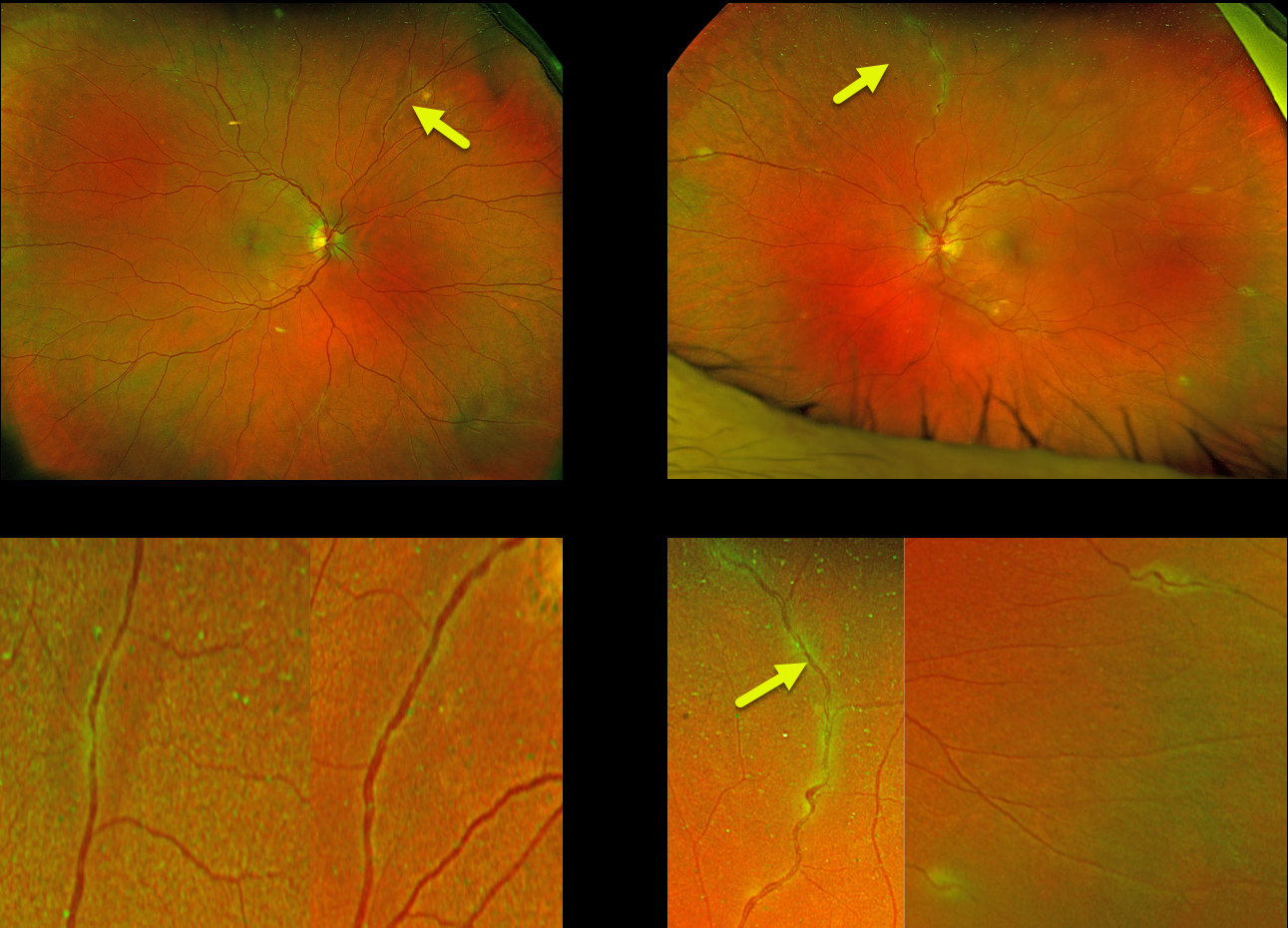
This 58YO female was referred for asymptomatic fundus changes. She gave a history of hypertension and COPD. Vision was 20/40 OU. Anterior segments were normal.
Optos color RG imaging shows bilateral multifocal areas of periphlebitis. Although sarcoid-specific bloodwork was negative (angiotensin converting enzyme, serum lysozyme, and soluble interleukin-2 receptor), chest CT showed multifocal, bilateral calcified and noncalcified pulmonary nodules throughout the lung parenchyma in addition to calcified mediastinal and hilar nodes. She was referred to a pulmonologist who agreed with our diagnosis of sarcoidosis. However, since there were no functional pulmonary deficits, he felt local treatment for the eye findings would be best. The patient will therefore be initially treated with a subtenon triamcinolone injection.
Learning Points:
Sarcoidosis, like syphilis, has a myriad of ocular presentations, including anterior, intermediate, and posterior uveitis, vasculitis, papillitis, and choroidal granuloma. Our patient’s segmental periphlebitis (‘candle wax drippings’) is virtually pathognomonic for sarcoidosis.
In our personal experience, as in our patient, there doesn’t appear to be a strong association between active systemic disease and ocular inflammation. Establishing the diagnosis of ocular sarcoid in the absence of a known systemic diagnosis is often difficult.
Sensitivity and specificity are highest for chest CT, and combined with elevated serum ACE and lymphopenia, are strongly suggestive of systemic sarcoidosis (Nieder and Sims, AJO 1019;206:149-153). Another recent publication found bilateral hilar lymphadenopathy on chest CT and sIL-2R as the most sensitive biomarkers for diagnosing systemic sarcoidosis (Japanese J Ophthalmology 2021;65:191-198).



