

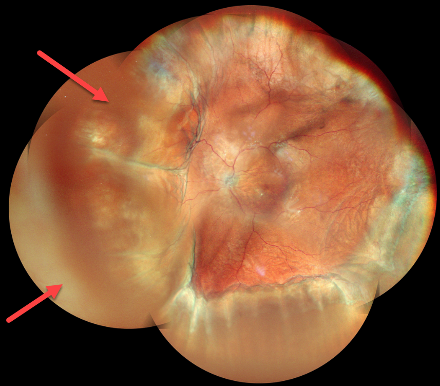
This 63YO male underwent glaucoma filtering surgery with mitomycin C one month earlier at an outside practice. Vision was 20/30, and intraocular pressure was 4. Montage color imaging shows extensive choroidal detachments. He was immediately lost to follow-up.
Learning Points:
A choroidal detachment is defined by the abnormal presence of fluid or blood in the suprachoroidal space. Serous choroidal detachments, also known as choroidal effusions, are a frequent complication of glaucoma surgery.
Several causative mechanisms are believed to be at play: hypotony allows fluid to accumulate in the suprachoroidal space, and inflammation increases the choroidal permeability. The detached ciliary body may also produce less aqueous humor, thus creating a self-perpetuating cycle. Often, observation is sufficient, but topical steroids and cycloplegics can be helpful in facilitating resolution.



