

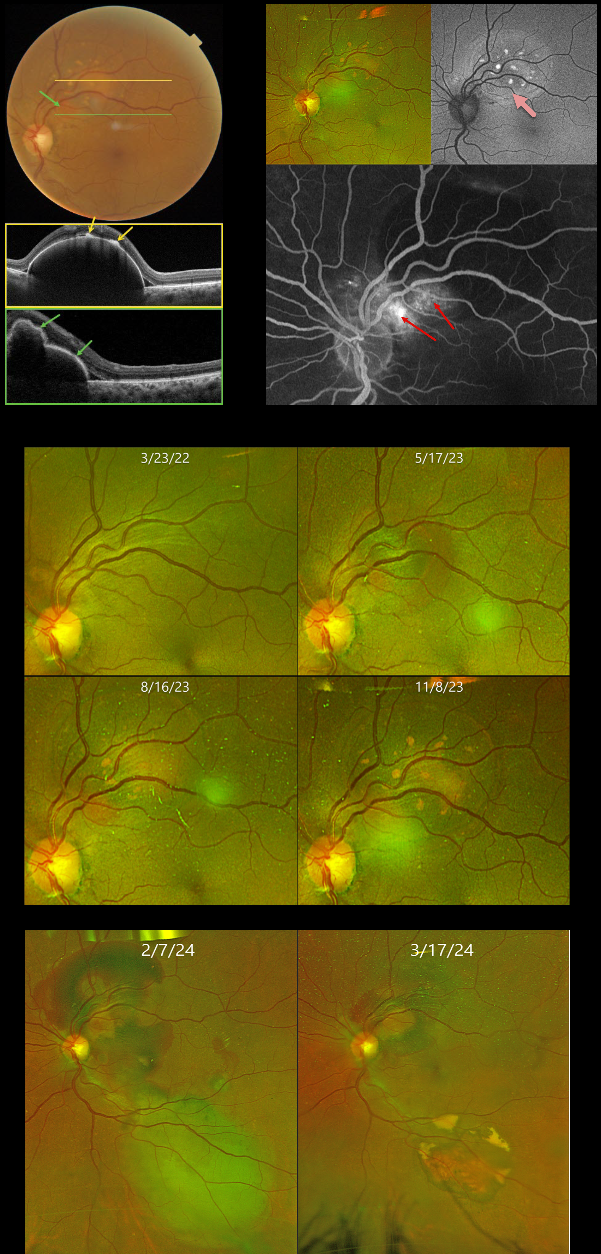
This 59YO female was initially referred to us on 3/23/22 with a 1-year history of vision loss in her right eye. Vision was counting fingers OD and 20/25 OS.
Over the past 10 months, a gradually enlarging RPE detachment developed in the left superonasal macula. On 11/8/23, Triton swept-source OCT confirms a highly elevated RPE detachment. The orange lipofuscin deposits seen on color imaging appear as hyperreflective lesions on OCT. Scanning through the reddish polyp shows a multi-lobulated elevation of the RPE. The lipofuscin deposits markedly hyper-autofluoresce. The large variably staining/leaking polyp creates a notch along the inferonasal edge of the PED.
Due to the patient’s history of a large macular hemorrhage resulting in counting fingers vision in her fellow eye, half-fluence photodynamic therapy (PDT) was applied to the polyp in her left eye. However, one month following PDT on 2/27/24, there was increased blood around the treated superior lesion, with new blood inferiorly.
Multiple full-fluence PDT applications were then applied to the old and new lesions. Six weeks later, the patient’s fundus was dramatically improved, with near-complete resolution of all blood and fluid. Vision was 20/20, and we will continue to follow her closely.
Learning Points:
Polypoidal choroidal vasculopathy (PCV) is a variant of type 1 macular neovascularization. PCV is likely distinct from age-related macular degeneration, occurring in a different population (more common among African Americans and Asians), with a relative lack of drusen and a thicker choroid. The sub-RPE vessels were classically described using indocyanine green angiography, although newer literature suggests that OCT is likely just as effective at diagnosing these vascular polyps (Permadi et al., Int J Retina Vitreous 2022;8;14).
Historically, these lesions were believed to be somewhat resistant to anti-VEGF monotherapy, often requiring combined treatment with photodynamic therapy. Newer data from the PLANET study suggest that Eylea monotherapy is also successful (AJO 2019;204:80-89).
Originally described by Gass (Retina 1984;4:205-220), a notched PED is a sign of an occult MNV. The notch represents a type 1 MNV, which tethers the overlying RPE from the surrounding more highly elevated PED. Hydrostatic leakage from the MNV may contribute to the larger PED.



