

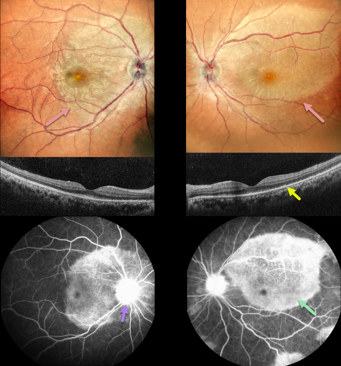
This 36YO female presented with a month of bilateral vision loss, which got worse over the prior 2 days. She also complained of 3 months of headaches and painful urination. Vision was 20/30 OD and 20/120 OS.
Multicolor imaging shows bilateral creamy placoid lesions. OCT scanning shows variable ellipsoid zone loss with small hyperreflective material above. Some vitreous cells are also noted. Fluorescein angiography shows late staining of these placoid lesions with optic nerve leakage.
Bloodwork was positive for syphilis, and she was referred to an infectious disease specialist for further management.
Learning Points:
Ocular manifestations of syphilis vary widely and include virtually all variations of anterior and posterior segment inflammation, including a recently described presentation as the multiple evanescent white dot syndrome phenotype (Russel et al Int Ophthalmol 2020;40:627-738). Hence, the reason it’s been dubbed the great imitator or masquerader.
One of the more common presentations is placoid chorioretinitis (Eandi et al, Retina 2012;32:1915-1941), which was evident in our patient’s multimodal imaging.



