

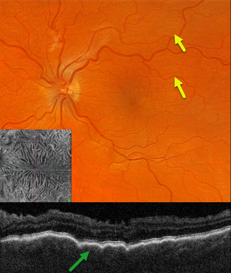
This 39YO male with a history of pigmentary glaucoma underwent goniotomy surgery by an outside practice in his left eye 3 weeks earlier. This eye has been hypotonus since, and vision was 20/150.
Optos color RG imaging shows faint diffuse chorioretinal folds throughout the posterior pole. Triton swept-source OCT B-scan shows a thickened choroid with overlying folds, and an irregularly rippled inner retina. En face OCT through the internal limiting membrane provides a unique perspective on inner retinal distortion.
Learning Points:
Hypotony maculopathy is characterized by low IOP and chorioretinal folds, most commonly following glaucoma filtration surgery. Optic nerve edema can also be seen due to decreased axoplasmic transport.
We expect our patient’s findings to improve once his intraocular pressure normalizes.



