

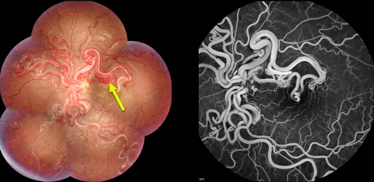
This healthy 16YO girl presented with a long history of poor vision in her left eye. Vision was 20/100 in her left eye.
Fundus photography of her left eye shows tangles of markedly dilated and tortuous vessels covering the posterior pole. Fluorescein angiography shows no leakage. Brain MRI revealed a cerebral hemangioma next to the optic chiasm.
Learning Points:
A retinal arteriovenous malformation (AVM) is a congenital connection between an artery and a vein without an intervening capillary network. These lesions appear along a spectrum ranging from small, isolated lesions to the large ‘bag of worms’ appearance as seen in our patient. These more extensive lesions are usually part of the Wyburn-Mason Syndrome, which includes vascular lesions in the eyes and midbrain. Retinal venous malformations, aka retinal macrovessels, are now thought to represent an AVM (Pichi et al JAMA Ophthalmol 2018;136:372-379).



