

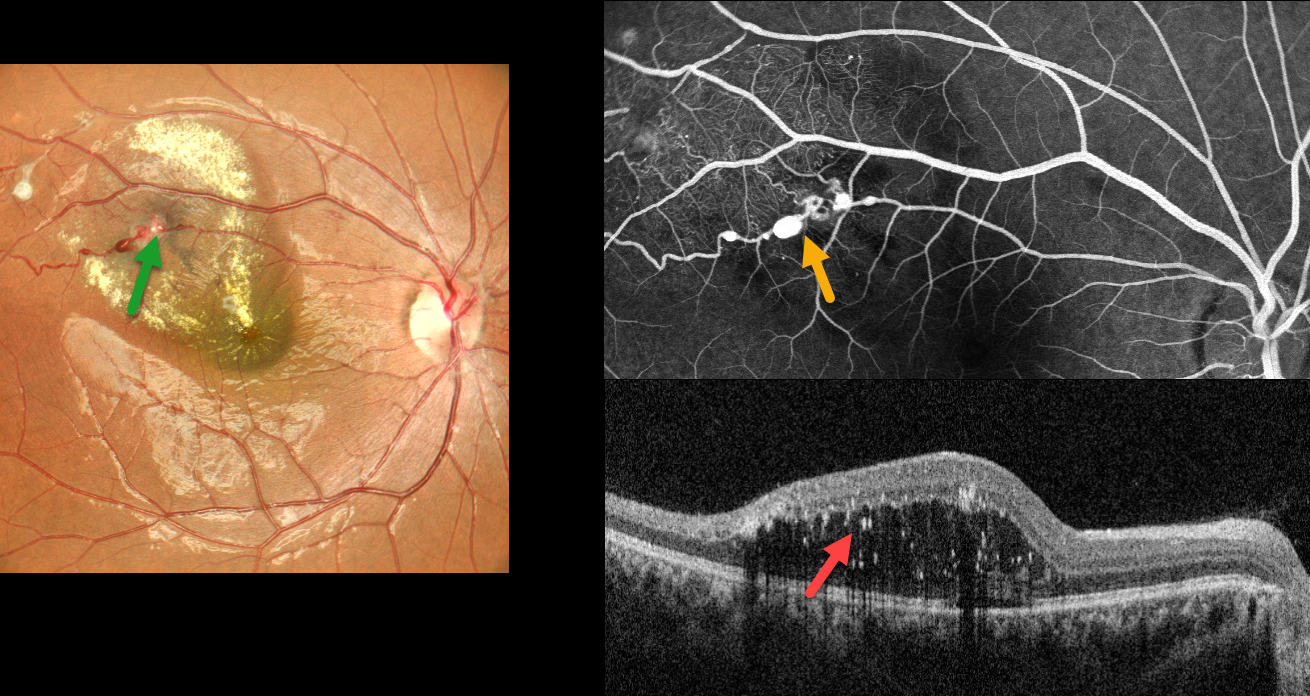
This healthy 19YO female presented with 1 week of blurred vision in her right eye. Vision was 20/80 OD and 20/20 in her normal OS.
Color imaging shows multiple superotemporal variably fibrosed macroaneurysms with surrounding lipid that extends towards the central macula. More subtle telangiectasia and lipid were seen in the superior periphery (not shown).
Fluorescein angiography shows multiple patent macroaneurysms along the superotemporal arteriole with more distal telangiectasia. OCT shows outer retinal fluid with numerous suspended hyperreflective lipid particles. Thermal laser followed by 3 monthly anti-VEGF injections was given.
Learning Points:
Described by George Coats in 1908, Coats disease is an idiopathic disorder that is usually unilateral in males. It is characterized by retinal vascular telangiectasia involving capillaries, arterioles, and venules. Patients can present with a wide variety of findings ranging from minimal exudation to total exudative retinal detachment. The leaking vessels are usually treated to prevent exudative complications.
Thermal laser photocoagulation is usually the initial modality, but anti-VEGF injections can also be used to stabilize the vascular leakage. Patients will often require multiple rounds of treatment. See Sigler et al for a review of the current management (Survey Ophthalmology 2014;59:30-46).
Dyskeratosis congenita or familial exudative vitreoretinopathy should be considered for patients with frank bilateral disease (Teixeira et al, Arch Ophthalmol 2008;126:134-135), in women with Coats-like findings (Johnson et al, Genetics 2009;30:181-184), or in patients with a family history of similar findings. Since our patient’s findings were unilateral and her nail beds were normal, we deferred genetic testing.



