

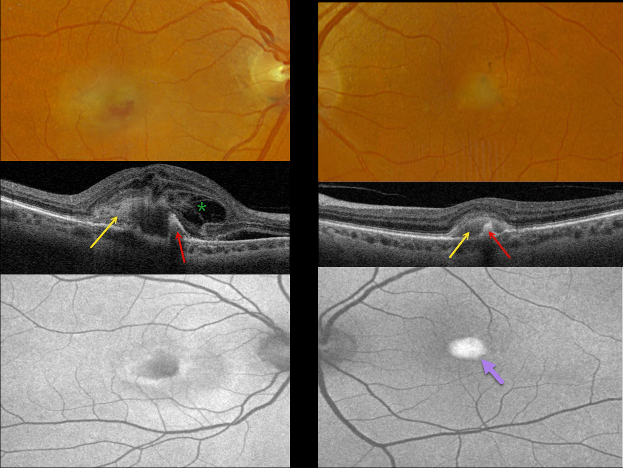
This 71YO female was referred for neovascular AMD. She noticed recent vision loss in her right eye and was fairly asymptomatic in her left eye. Vision was counting fingers OD and 20/80 OS.
Optos color RGB imaging shows somewhat turbid, yellowish foveal fluid OD and a vitelliform lesion OS. Triton swept-source OCT shows hyperreflective subretinal vitelliform material and hyperreflectivity from a subretinal pigment clump. Subretinal fluid and a complex pattern of loculated pockets of intraretinal fluid are noted OD.
On Optos fundus autofluorescence (FAF), the lesion shows central hypo-FAF and peripheral hyper-FAF OD, and marked hyper-FAF OS.
Anti-VEGF therapy was started for macular neovascularization (MNV) in her right eye.
Learning Points:
Best disease is associated with a mutation in the BEST1 gene, which encodes bestrophin-1. Bestrophin-1, a calcium-activated chloride channel, is primarily found in the basolateral plasma membrane of the RPE.
BEST1 mutations cause a variety of phenotypes, including adult-onset foveomacular vitelliform dystrophy (as in our patient), best vitelliform macular dystrophy, autosomal recessive bestrophinopathy, and autosomal dominant vitreoretinochoroidopathy.
Although our patient had a Best phenotype, genetic testing revealed uncertain significant heterozygous variants SAG, SLC7A14, and VCAN.
In our experience, MNV is rarely associated with Best lesions, although Miyagi et al identified MNV in 18% of eyes in their retrospective Asian population (Graefe’s 2022;260:1125-1137).



