

This 43YO female presented to our office with recent vague symptoms of vision loss OD. She was followed elsewhere since 2006 for a carcinoid lesion metastatic to the right choroid. Her right bronchial carcinoid tumor was treated earlier that year with a right pneumonectomy without systemic treatment. Initial observation of the choroidal lesion was recommended, but she was almost immediately lost to follow-up for 13 years.
She returned to the outside practice in 2019, and the choroidal lesion showed slight growth with mild new fluid. She was again lost to follow-up until June 2021, with continued growth and increased fluid. Photodynamic therapy was applied on 6/8/21 and 9/21/21 with subsequent tumor shrinkage and resolution of the fluid.
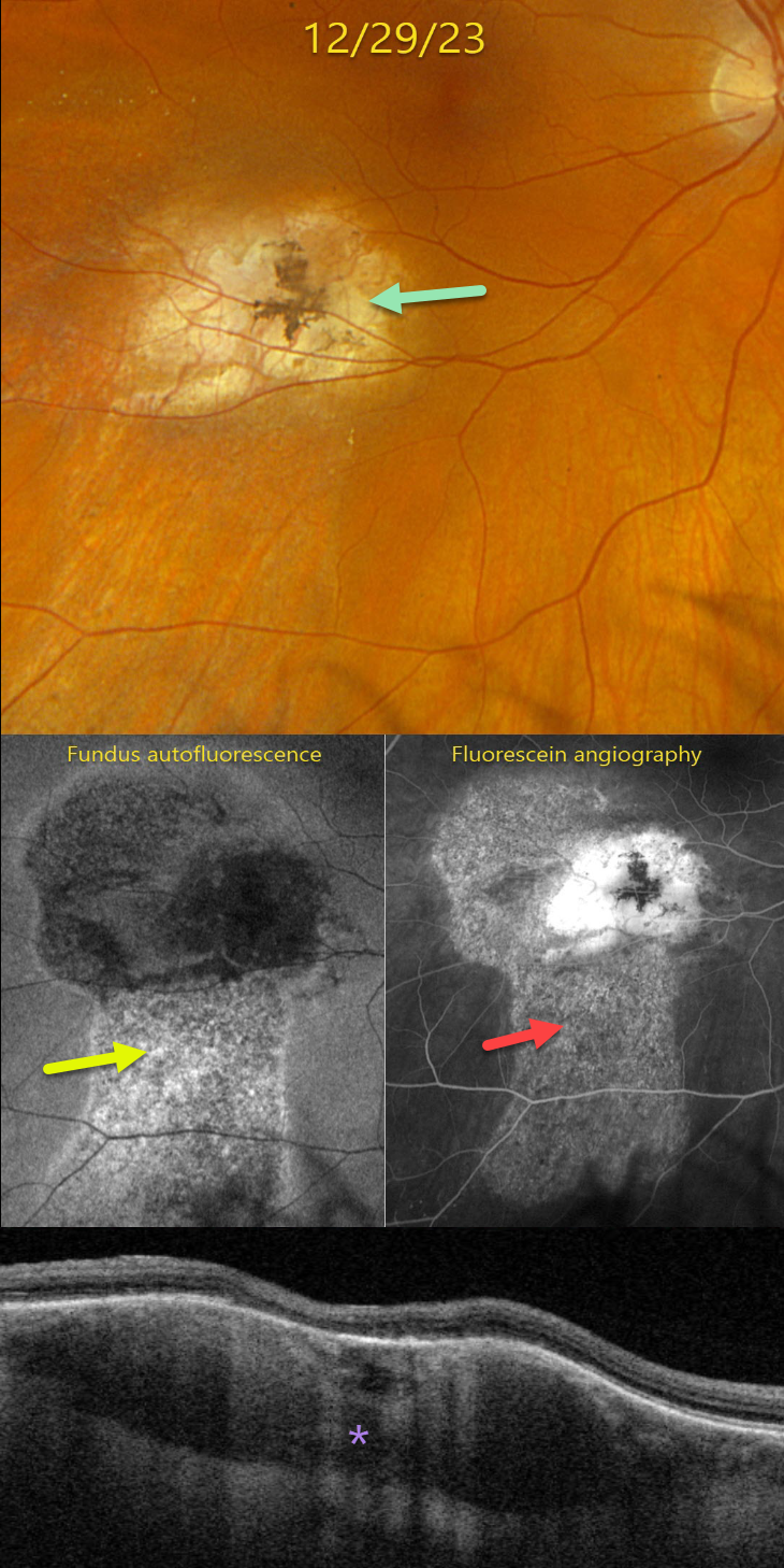
On 12/29/23, vision was 20/25. Optos color RGB imaging shows an atrophic, variably pigmented, lumpy choroidal lesion in the inferotemporal macula.
Fundus autofluorescence (FAF) shows a hyper-FAF gutter extending inferiorly from the lesion. This gutter is well-visualized on fluorescein angiography, with staining of the inactive tumor.
Triton swept-source OCT shows a variably thickened and mostly hyporeflective choroidal lesion with overlying outer retinal atrophy. These findings were all stable compared to images seen 1 year earlier at the outside practice. We plan to follow her twice a year.
Learning Points:
Although uveal melanoma is the most common primary intraocular malignancy, metastatic choroidal tumors are the most common intraocular malignancies. Lung and breast cancer are the most common sources. These lesions can be treated with systemic chemotherapy and are also usually very responsive to external beam radiation.
Neuroendocrine (carcinoid) tumors are usually low-grade malignancies that arise in the bronchopulmonary system or the gastrointestinal or urogenital tracts. In a recent review of over 1100 patients referred to the Wills ocular oncology service over a 43-year period, pulmonary carcinoid accounted for 2% of cases (Shields et al, Retina 2020;40:204-213). These lesions have a characteristic orange appearance, in contrast to most other choroidal metastases, which are usually creamy yellow (Shields et al, BJO 1996;80:852-853).



