

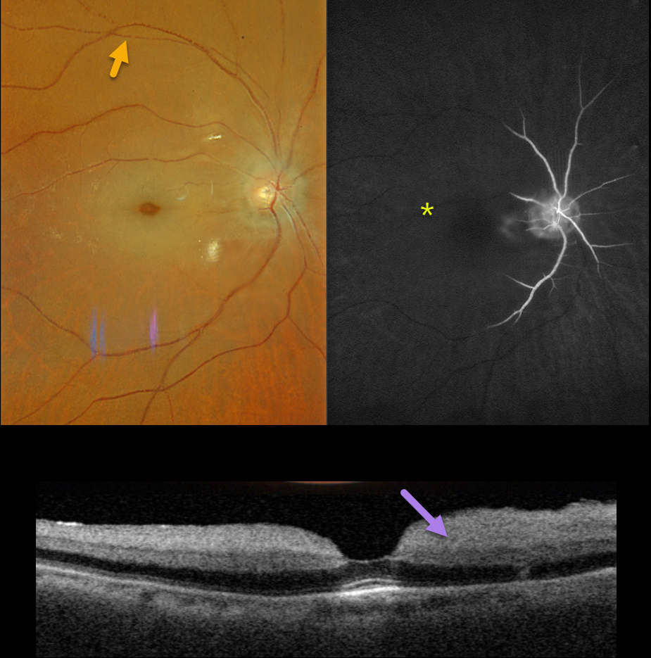
This 89YO male presented with 2 days of acute and severe vision loss in his right eye. Vision was no light perception.
Optos color RGB imaging shows an acute central retinal artery occlusion (CRAO) with a cherry-red spot and a stagnant blood column, best seen superiorly as box-carring of the column.
Fluorescein angiography shows profound ischemia, with no vascular filling beyond the peripapillary region.
Triton swept-source OCT shows a diffusely slightly thickened and markedly hyperreflective inner retina. He was immediately referred to the nearest stroke center.
Learning Points:
Patients with acute retinal ischemia (defined as transient monocular vision loss, acute branch retinal artery occlusion, or acute CRAO) require emergent referral to a stroke center. This is especially urgent with an acute CRAO since about 75% of patients have already developed a recent stroke.
If the patient presents immediately following the occlusion, measures to acutely lower the intraocular pressure to move the causative embolus downstream can be performed, including ocular massage, paracentesis, and breathing into a bag.
Unfortunately, most patients present outside this window (Shah et al, Ophthalmology Retina 2023;7:527-531) and even then, these measures are usually ineffective.



