

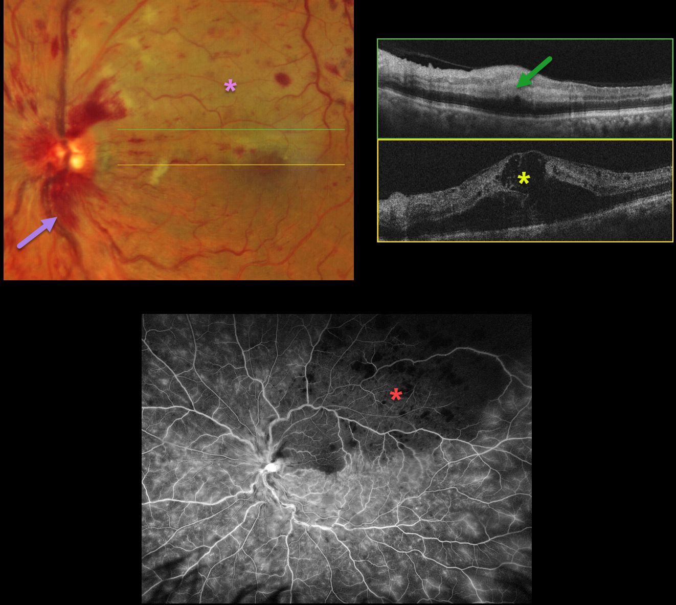
This 66YO male presented with sudden vision loss in his left eye. Vision was 20/30 in his normal right eye and counting fingers in his left eye.
Optos color RG imaging shows a central retinal vein occlusion (CRVO) with mild retinal hemorrhages in all quadrants, except for more moderate hemorrhages superotemporally. A secondary superior branch retinal artery occlusion (BRAO) bisects the fovea.
OCT scanning shows marked central edema, with hyperreflective and thickened inner retina within the BRAO. Fluorescein angiography shows a well-perfused CRVO, except for delayed arterial filling and ischemia within the BRAO.
Learning Points:
Some patients with CRVO will develop a secondary non-embolic BRAO caused by compression from optic nerve edema. However, our patient’s optic nerve did not appear that swollen. Our patient was started on anti-VEGF therapy for the severe macular edema.



