

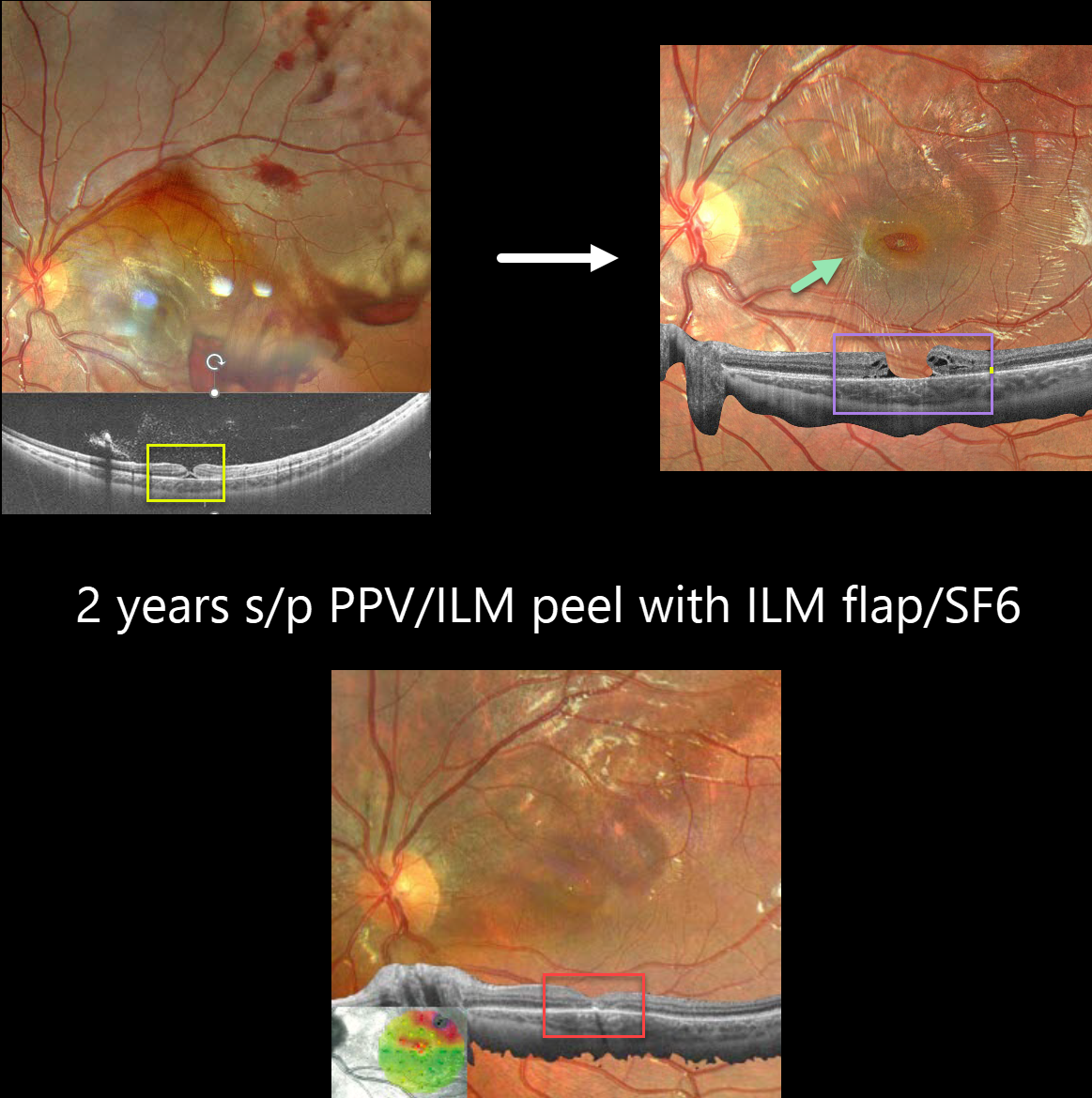
This 10YO boy was referred for vision loss immediately after getting hit in his left eye by a soccer ball 7 days earlier. Vision was 20/70.
Fundus photography shows scattered intra- and preretinal hemorrhages with a large confluent area of commotio retina superiorly and temporally.
Widefield OCT shows a partial-thickness macular hole with the external retina intact but elevated. Three months later, vision was 20/100, and an irregular, full-thickness macular hole with surrounding macular pucker was seen.
25-gauge vitrectomy with ILM peeling, ILM flap, SF6 gas tamponade, and 3 days face-down positioning was performed. Two months later, vision improved to 20/40 with complete closure of the hole. Two years later, vision was 20/25.
Learning Points:
This patient illustrates several sequelae of blunt trauma, including retinal/preretinal blood, commotio retinae, and macular hole formation. Since fresh traumatic holes can spontaneously flatten, especially in pediatric cases (Liu et al, AJO 2020;210:174-183), an initial period of observation is recommended before considering vitrectomy.
In our patient’s case, the hole did not resolve, and surgery provided good anatomic and long-lasting visual outcomes.



