

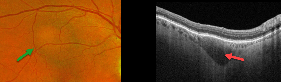
This 84YO male was referred for an asymptomatic macular lesion in his right eye. He was a +6 diopter hyperope. Vision was 20/30 in his right eye and 20/30 in his left eye.
Optos color RG imaging shows a subretinal, mostly depigmented lesion in the superior macula. Triton swept-source OCT shows a hyporeflective outer choroidal lesion that bows posteriorly. The choroid surrounding this lesion is relatively hyperreflective and disorganized. Observation was recommended.
Learning Points:
Initially described by Freund et al (Arch Ophthalmol 2003;121:197-204), intrachoroidal cavitations are most commonly found in high myopes as a peripapillary yellow-orange subretinal lesion. Our case is quite unique, given the location and occurrence in a hyperope.



