

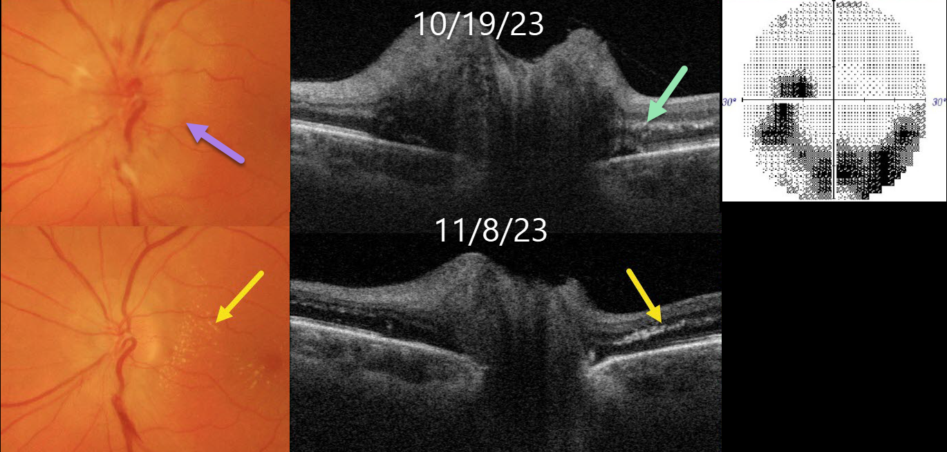
This healthy 56YO female presented with 1-2 weeks of blurred vision in her left eye. Vision was 20/25 in her normal OD and 20/100 OS.
Color imaging shows a diffusely swollen left nerve. Optic nerve Triton swept-source OCT B-scan confirms the diffuse retinal nerve fiber layer edema. A few dots of subclinical hyperreflective lipid are noted in the outer plexiform layer. 30-2 visual field testing shows an inferior unilateral arcuate scotoma extending from the blind spot.
Extensive blood work and chest X-ray were negative except for a positive QuantiFERON-TB Gold. Infectious disease consultation led to the diagnosis of latent tuberculosis, and she was started on systemic anti-tuberculous therapy.
Over the following several weeks, as the optic nerve edema resolved, the papillomacular outer plexiform layer lipid became more prominent.
Learning Points:
The diagnosis for a unilateral swollen nerve is extensive. However, it becomes much smaller when associated with acute inner retinal ischemia (not seen in our case) and later lipid exudation within Henle’s layer (neuroretinitis), including cat scratch (Bartonella), Lyme disease, and syphilis. When no underlying cause is found, the entity is called Leber’s idiopathic stellate neuroretinitis.
Tuberculosis is a rare cause of neuroretinitis (Stechschulte et al, J Neuroophthalmology 1999;19:201-204). It is unclear whether our patient’s findings are idiopathic or related to her latent tuberculosis.



