

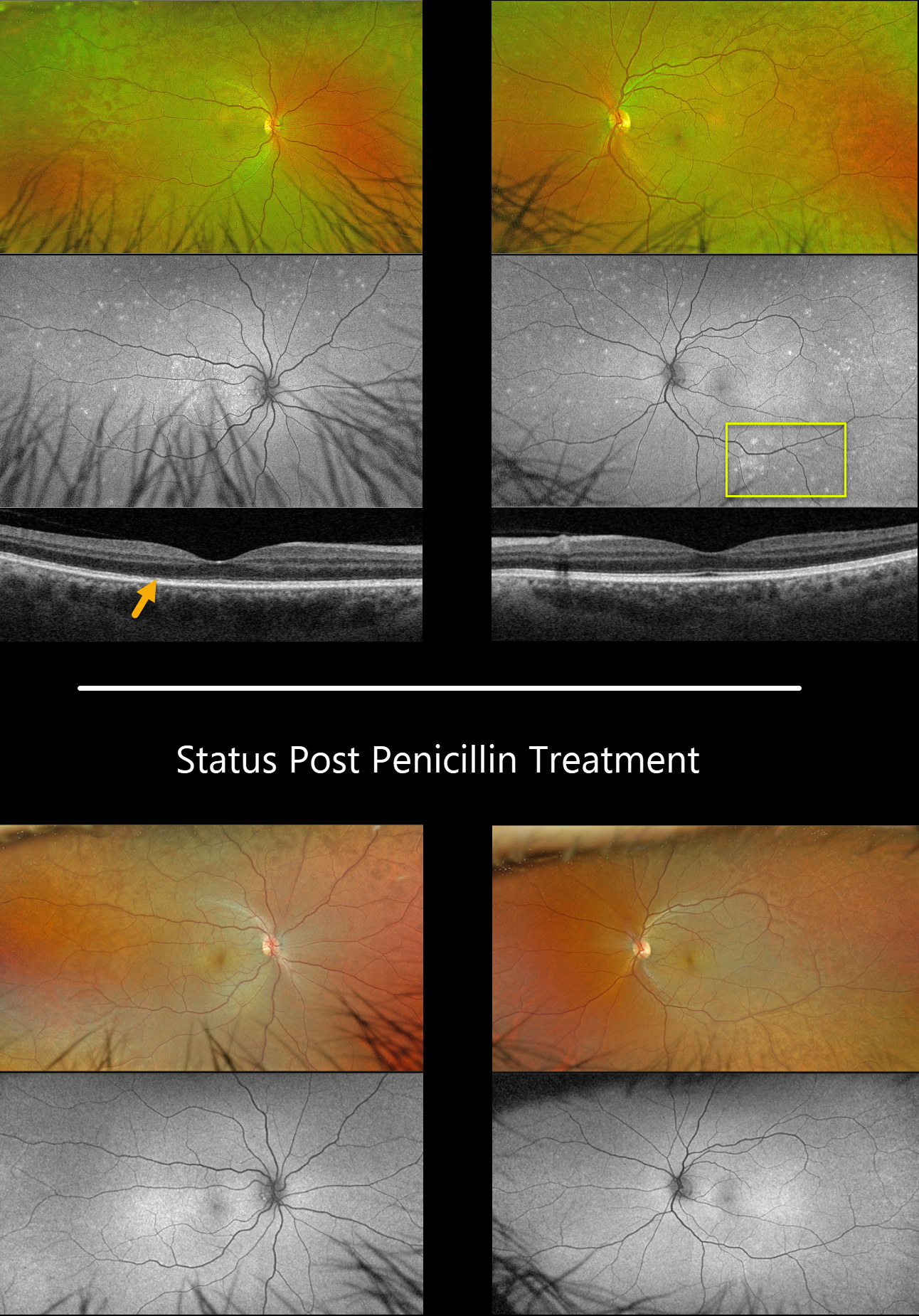
This 57YO male presented with 3 weeks of blurred vision in his right eye. Vision was 20/40 OD and 20/25 OS.
Optos color RG imaging shows bilateral plaques of whitish deep retinal discoloration. Fundus autofluorescence (FAF) shows multifocal MEWDS-like hyper-FAF spots scattered throughout each fundus. Triton swept-source OCT shows an indistinct temporal foveal ellipsoid zone OD and a fairly normal B-scan OS.
RPR testing was positive, and the patient was started on parenteral penicillin treatment. The autofluorescent findings dramatically resolved seven weeks later.
Learning Points:
Ocular manifestations of syphilis vary widely and include virtually all variations of anterior and posterior segment inflammation, including a recently described presentation as the multiple evanescent white dot syndrome phenotype (Russel et al Int Ophthalmol 2020;40:627-738). Hence, the reason it’s been dubbed the great imitator or masquerader.



