

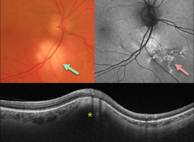
This 73YO male presented with this asymptomatic amelanotic lesion inferior to the right optic nerve. Optos fundus autofluorescence (FAF) shows overlying hyper-FAF with some dendritic radiating lines of hyper- and hypo-FAF extending inferonasally.
Triton swept-source OCT scanning through this region shows hyperreflective scleral thickening, which compresses the overlying choroid and elevates the retina.
Learning Points:
Focal scleral nodule, previously known as solitary idiopathic choroiditis (SIC) and unifocal helioid choroiditis, presents as a single, yellow-white, elevated nodular, avascular subretinal lesion. On OCT, it arises from the inner sclera, causing marked compression of the choroid.
For the largest series to date and the rationale behind its renaming, see Fung et al, Ophthalmology 2020;127:1567-1577.



