

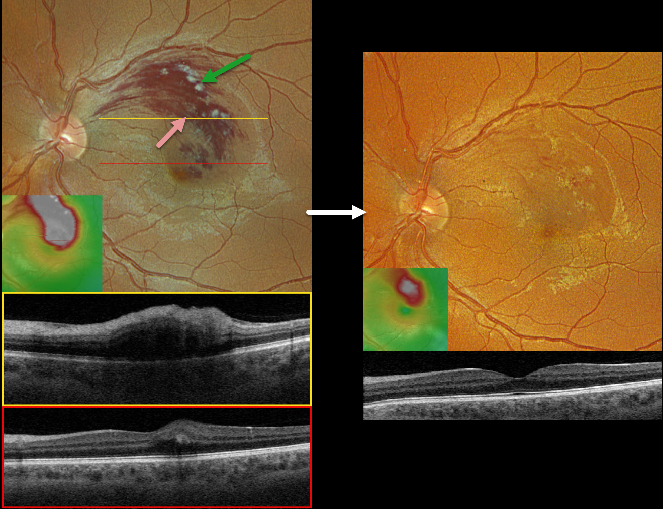
This healthy 33YO male presented with a 1-week history of vision loss in his left eye. Vision was 20/20 in his normal right eye and 20/60 in his left eye.
Optos color RGB imaging shows a superior macular branch retinal vein occlusion (BRVO) with relatively confluent inner retinal hemorrhages and superiorly located nerve fiber layer infarcts. Retinal fluid extends through the fovea.
Triton swept-source OCT through the superior macula shows retinal thickening and inner hyperreflectivity from the blood. Scanning through the macular center shows very shallow central edema. The macular cube shows the extent of the retinal thickening.
Following several monthly Avastin injections, vision improved to 20/30, and the macula was virtually free of fluid.
Learning Points:
Macular grid laser photocoagulation (Branch Vein Occlusion Study Group, AJO 1984;96:271-282) was the mainstay for treating persistent vision loss due to BRVO macular edema until the advent of intravitreal steroid and anti-VEGF therapy in the early-to-mid 2000s.
Although we were initially ‘WOW’ed by the near immediate and dramatic edema improvement that was never seen with thermal laser, the SCORE Study Research Group in 2009 suggested that grid laser should “remain the benchmark against which other treatments are compared” due to similar vision between intravitreal triamcinolone and laser but with fewer laser adverse events (Arch Ophthalmol 2009;127:1115-1128).
Intravitreal anti-VEGF injections have subsequently become the standard of care for initial therapy. A recent systematic review and meta-analysis showed that a treat-and-extend protocol is as effective as monthly and PRN regimens (OSLI Retina 2023;54:131-138).
Adding macular laser probably does not reduce the treatment burden or further improve vision (Ojima et al, Retina 2023;43:999-1004).



