

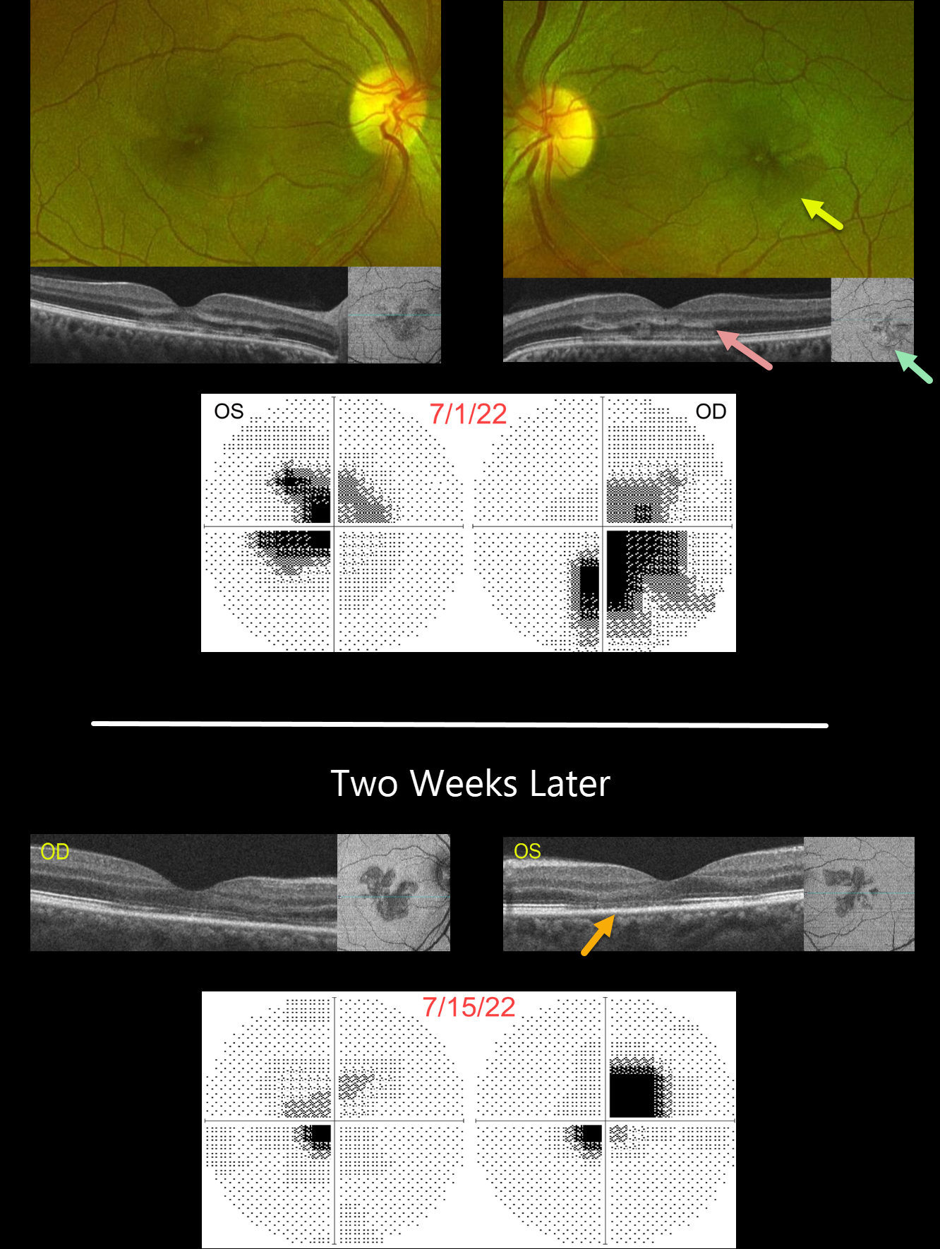
This 41YO female presented with recent bilateral paracentral scotomas and blurred vision. There was no prior ocular or past medical history. Vision was 20/50 OD and 20/100 OS.
Optos color RG imaging shows faint wedge-shaped lesions radiating outwards from each macular center. OCT B-scanning shows variable patchy hyperreflectivity extending from the outer plexiform layer into the interdigitation zone. OCT en face imaging through the outer retina shows hyporeflective wedge lesions. Central 10-2 visual field testing shows bilateral paracentral scotomas.
Two weeks later, vision spontaneously improved to 20/60 OD and 20/25 OS. On OCT B-scanning, the outer retinal hyperreflective lesions are now replaced by secondary thinning with more prominent en face wedge lesions. The visual field scotomas are also improving.
Learning Points:
This case highlights many characteristic features of acute macular neuroretinopathy (AMN), an idiopathic disorder likely caused by compromise of the deep retinal capillary plexus.
Traditionally, the pathognomonic wedge-shaped AMN lesions are best seen with near-infrared reflectance, although we also find them easily imaged with en face OCT. With resolution, the acute lesions cause secondary outer retinal thinning.
Most patients are white, non-Latino females with a median age of 30 years. The most common associations are a flu-like prodrome and oral contraceptive use. Presenting acuity is usually good, coinciding with sparing of the sub-foveal areas. Roughly half of AMN cases are bilateral. There is typically moderate visual improvement over several months without treatment.



