

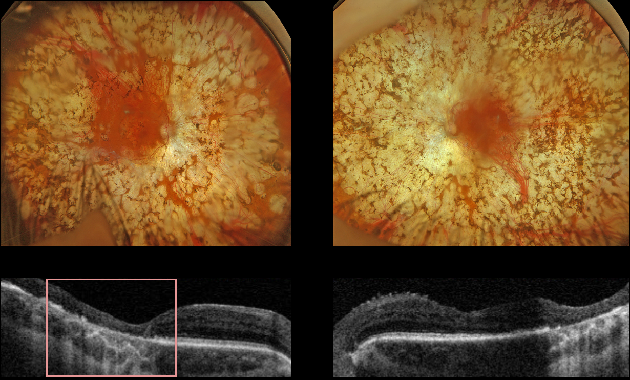
This 53YO female with type 1 diabetes received bilateral panretinal photocoagulation (PRP) for proliferative diabetic retinopathy (PDR) about 30 years previously from an outside practice. She presented for her yearly retinopathy examination, and vision was stable at 20/200 OD and 20/30 OS.
Optos color RGB imaging shows bilateral confluent PRP scarring extending from within the arcades into the peripheries. Triton swept-source OCT of the right eye shows outer retinal atrophy extending through the foveal center from confluent macular laser scarring, with more temporal outer retinal atrophy in the left eye from confluent posterior PRP scarring.
Learning Points:
PRP should begin at least 1 disc diameter from the optic nerve and outside the major temporal arcades. Treatment is performed with a wide-angle lens or an indirect ophthalmoscope, and should be applied from the posterior retina towards the periphery to avoid accidental macular burns. The laser spots should be 200-500 microns in size at the retina, be spaced about one burn-width apart, and should extend as far anteriorly as possible.
The laser performed on this patient is wrong on so many levels, including spots extending into the macula, touching the nerve, and confluent elsewhere. The laser surgeon essentially gave this patient severe iatrogenic retinitis pigmentosa.
Although possibly appropriate before the advent of modern vitrectomy surgery and anti-VEGF therapy, this degree of laser should never be seen in today’s patients.



