

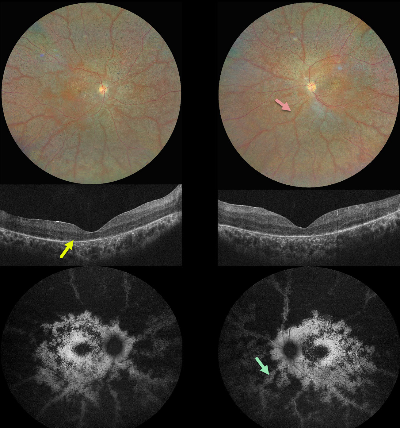
This 16YO male complained of night blindness for a few years. There was no family history of eye disease. Vision was 20/200 OU.
Color photography shows bilateral findings consistent with retinitis pigmentosa (RP), including diffuse chorioretinal scarring, intraretinal pigment migration, and narrow retinal vessels. The retinal tissue beneath and immediately surrounding the arterioles is relatively spared, and this is most dramatically seen on fundus autofluorescence. OCT scanning shows variable loss of the outer retinal bands and RPE. Our patient could not afford genetic testing and was immediately lost to follow-up.
Learning Points:
Preserved para-arteriole retinal pigment epithelium (PPRPE) in retinitis pigmentosa, originally described by Heckenlively (Br J Ophthalmol 1982;66:26-30), has the unique phenotype, as seen in our patient, of diffuse RP degeneration with relative sparing of the RPE beneath the retinal arterioles. It was later found to be caused by a mutation in the crumbs cell polarity complex component 1 (CRB1) gene (den Hollander et al, Nature Genetics 1999;23:217-221). CRB1 is part of a group of proteins that help determine the structure and orientation of photoreceptors. It may also be involved in forming connections between different types of retinal cells.



