

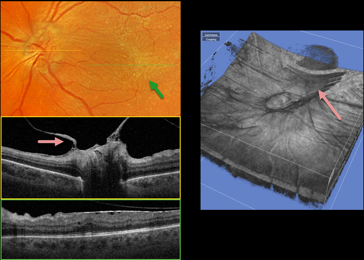
This 58YO male with type 2 diabetes has long-involuted proliferative diabetic retinopathy (PDR) following full panretinal photocoagulation (PRP). He has no visual complaints, and vision is 20/50 OS.
Optos color RGB imaging shows a diffuse macular pucker with striae extending nasally towards the nerve and temporally into the distal macula.
A condensation of elevated fibrosed tissue, representing a partially detached Weiss’ ring, surrounds the optic nerve. This traction is clearly evident in Triton swept-source OCT (image 1, middle) and in the 3D reconstruction (image 2). A moderate epiretinal membrane extends throughout the macula (image 1, bottom).
Observation was recommended, and vitrectomy with membrane peeling will be performed if the patient becomes more symptomatic.



