

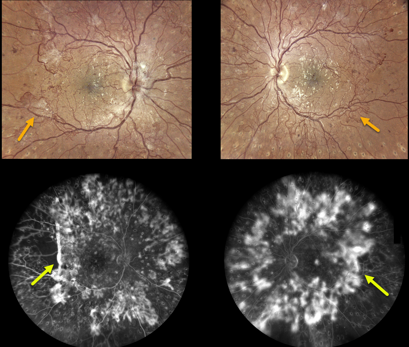
This 28YO female with type 1 diabetes recently received bilateral full panretinal photocoagulation (PRP), completing treatment several months earlier.
Despite this, active neovascularization encircles the major arcades, extending into the midperipheries, clinically and angiographically. She was visually asymptomatic with vision of 20/25 OD and 20/30 OS. There was no edema or traction on OCT (not shown).
After extensive discussion with our patient, we decided against fill-in PRP or anti-VEGF therapy for fear of possibly causing contraction of the neovascularization and traction detachment. We will follow her closely and recommend vitrectomy if vitreous hemorrhage or traction develops.
What would you do?
Learning Points:
Before PRP, patients with proliferative diabetic retinopathy (PDR) routinely went blind, and one of the treatments actually included pituitary ablation (for an amazing historical perspective on diabetic laser photocoagulation, see Goldberg and Jampol, Ophthalmology 1987;94:741-746).
Following PRP, the retinal neovascularization usually morphs from active (tight small red vascular networks) into involuted (residual larger vessels, fibrosed tissue, or occasionally complete vessel disappearance) vasculature.
Once considered a “one and done” treatment, more recent studies show that patients may still need additional laser, anti-VEGF injections, or vitrectomy (see Gonzalez et al, Ophthalmology 2021;128:1448-1457).



