

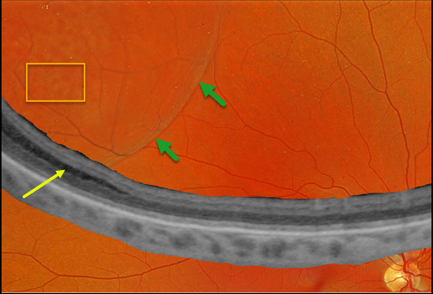
This 46YO female was referred for a possible retinal detachment. She had no visual complaints. Vision was 20/20 OD and 20/20 in her normal OS.
Optos color RGB imaging shows a bullous superotemporal retinoschisis. Numerous inner-layer holes are seen. Optos Silverstone ultrawidefield OCT shows splitting of the outer plexiform layer, confirming a schisis.
Learning Points:
Degenerative retinoschisis is an age-related splitting of the outer plexiform layer, most commonly located inferotemporally. It is sometimes difficult to differentiate a schisis from a rhegmatogenous detachment, and ultrawidefield OCT imaging, as in this case, is an amazing diagnostic tool.
Patients can rarely develop a combined schisis rhegmatogenous retinal detachment (RRD) when liquid vitreous accesses the subretinal space through both an inner-layer and an outer-layer hole. We will follow up with our patient yearly.



