

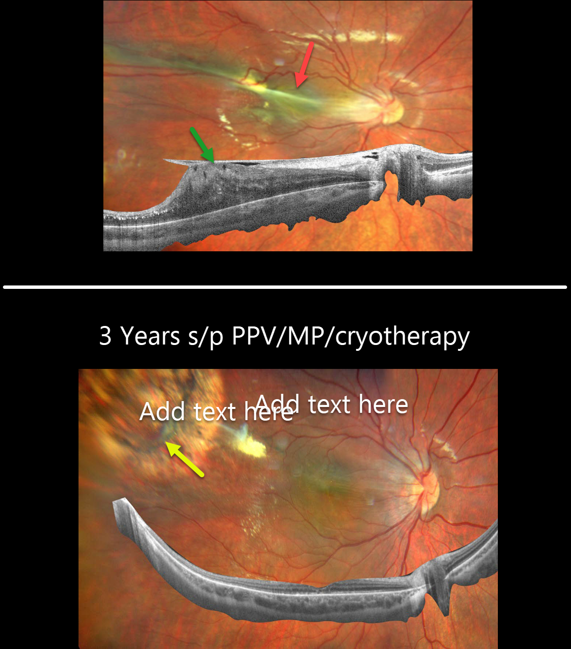
This otherwise healthy 8YO male presented with a chronic history of vision loss in his right eye. Vision was counting fingers OD and 20/25 OS.
Fundus photography shows dragging of the optic nerve and macula temporally towards an area of dilated, anomalous retinal vessels. Widefield OCT of the right shows an adherent epiretinal membrane elevating and dragging the nerve and macula temporally.
Pars plana vitrectomy with membrane peeling and cryotherapy of the area with anomalous vessels was performed. Three years later, vision improved to 20/25 OD. The macula is clinically and on OCT completely flat, with a dry cryotherapy scar noted in the superotemporal midperiphery.
This Eyecare Clinic (in Brescia, Italy) case was submitted by Barbara Parolini, Veronika Matello, Giulia Freschi, and Roberta Penzani.
Learning Points:
Originally described by Criswick and Schepens (AJO 1969;58:578-594), familial exudative vitreoretinopathy (FEVR) can be inherited as an autosomal-dominant, recessive, or X-linked trait with high penetrance and variable expressivity. There are numerous genes associated with FEVR, including LRP5, FZD4, NDP, TSPAN12, ZNF408, CTNNB1 and KIF11 [Tao et al, Invest Ophthalmol Vis Sci 2021;62(15);4].
FEVR is characterized by peripheral temporal retinal avascularity, lipid exudation, neovascularization, tractional retinal detachment, and temporal dragging of the macula and retinal vessels. These findings are somewhat similar to those found with retinopathy of prematurity (ROP).



