

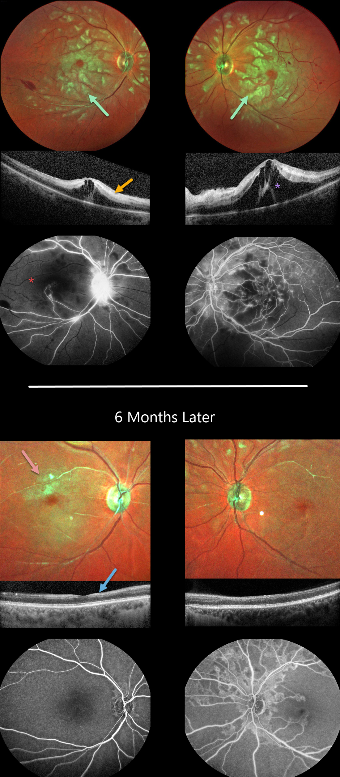
This previously healthy 29YO female presented to the emergency room with acute bilateral vision loss. Vision was counting fingers OU.
Color imaging shows bilateral white patches of inner and mid-retinal ischemia, white narrowed arterioles, and a few inner retinal hemorrhages.
OCT scanning shows variable hyperreflectivity within the inner and mid-retinal layers and cystoid edema. Fluorescein angiography confirms profound retinal ischemia, particularly in the right eye.
Extensive medical workup resulted in a diagnosis of systemic lupus erythematosus (SLE). Rheumatology started systemic rituximab. Bilateral intravitreal dexamethasone followed by intravitreal Eylea was given for the macular edema.
Six months later, although her vision remained at counting fingers OU, she was functioning better and learning to adapt to a new way of living. She was on prednisone 10mg daily and Eylea OU q12 weeks.
Although the acute patches of white retinal ischemia resolved, each fundus is clinically and angiographically markedly ischemic. OCT scanning shows no edema, although there is relative disorganization and thinning of the inner retinal layers.
Learning Points:
Purtscher retinopathy was first described by Otmar Purtscher in 1910. Although originally described in a man who fell from a tree with cranial trauma, these white patches of retinal ischemia and hemorrhages are found in numerous other etiologies, including pancreatitis, chest trauma, and collagen vascular diseases.
The findings are called Purtscher retinopathy when due to trauma, and Purtscher-like retinopathy when seen from other causes. SLE-associated retinopathy more typically appears as hypertensive retinopathy (bilateral nerve fiber layer infarcts and retinal hemorrhages), often in patients with cerebral vasculitis. However, our patient’s blood pressure was not elevated, and there were no signs of central nervous system involvement.
The underlying cause of Purtscher retinopathy is unknown, but it is thought to result from leukoembolization mediated by complement activation. The areas of ischemia include more superficial nerve fiber layer infarcts (cotton wool spots) and deeper ischemia involving the middle retinal layers.
This middle retinal ischemia, paracentral middle maculopathy (PAMM), is a recently-described phenotype found as an idiopathic entity or in other ocular disorders, including Purtscher, diabetic retinopathy, retinal artery and retinal vein occlusions, and acute macular neuroretinopathy (see Retina 2015;35:1921-1930).



