

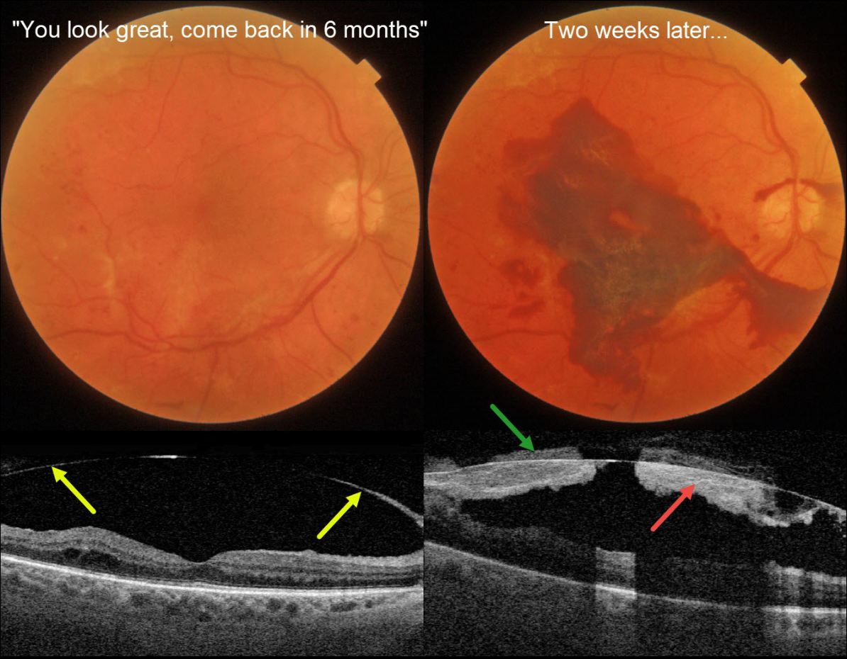
This 71YO diabetic male was examined over a year following completion of panretinal photocoagulation (PRP) for proliferative diabetic retinopathy (PDR). Vision was 20/50.
Triton color photography shows no macular edema, and swept-source OCT reveals a detached hyaloid elevated over the macula. He was told that everything looked great and to come back in 6 months. Famous last words…
Two weeks later, he presented with acute vision loss in this eye. Vision was counting fingers from a fresh macular subhyaloid hemorrhage. Both subhyaloid and prehyaloid blood are noted on Triton swept-source OCT.
An intravitreal Avastin injection was given, and vitrectomy will be recommended if the blood fails to clear.
Learning Points:
Although PRP is often a ‘one and done’ procedure, this is not always the case (Gonzalez et al, Ophthalmology 2021;128:1448-1457), and patients therefore need continued monitoring. Although our patient appeared perfect 2 weeks earlier, this case reminds us to remain humble in the presence of the retina and that even an involuted PDR requires lifelong follow-up.



