

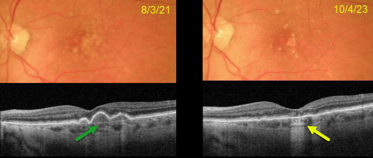
This 77YO female presented on 8/3/21 with 20/30 vision in her left eye. Optos color RG imaging shows confluent central soft drusen, which are confirmed on Triton swept-source OCT.
On 10/4/23, vision was 20/25. The central drusen flattened with secondary drusenoid atrophy. Continued observation was recommended.
Learning Points:
Drusen commonly remodel over time and their collapse, as in our patient, can lead to drusenoid atrophy of the RPE (Filho et al, Retina 2013;33;1558-1566). Vision may or may not decrease following regression of drusen.
In our experience, drusenoid atrophy remains stable, unlike primary age-related macular atrophy, whose radius enlarges linearly over time (Shen et al, Ophthalmology Retina 2020;4;899-910). We therefore do not consider intravitreal complement inhibitors for these patients.



