

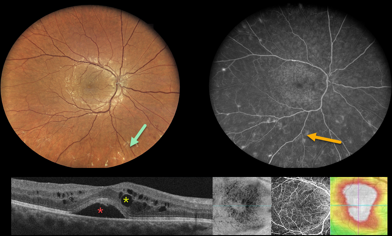
This 38YO diabetic female was referred for diabetic retinal changes. Vision was 20/50 OD and 20/40 OS. Multimodal imaging shows the often subtle proliferative changes with various perspectives of her center-involved diabetic macular edema (CI-DME).
Widefield color imaging shows minimal retinal hemorrhages with a foveal cyst. Upon closer inspection, subtly fibrosed retinal neovascularization is noted, particularly inferiorly. Fluorescein angiography shows numerous buds of retinal neovascularization outside the macula.
Multiple OCT images are shown below, including a B-scan, which shows subfoveal fluid with cysts in both the inner and outer nuclear layers. En face imaging shows variably sized radiating cysts. OCT angiography is fairly normal with a preserved foveal avascular zone. Finally, a thickness map shows diffuse thickening.
Learning Points:
These presentations can be very humbling for us clinicians and remind us of the importance of peripheral retinal examination and ultra-widefield imaging with fluorescein angiography in screening for proliferative disease. Widefield OCT angiography may also prove to be an equally effective yet less invasive screening tool (Al-Khersan et al., AJO 2021;224:292-300).



