

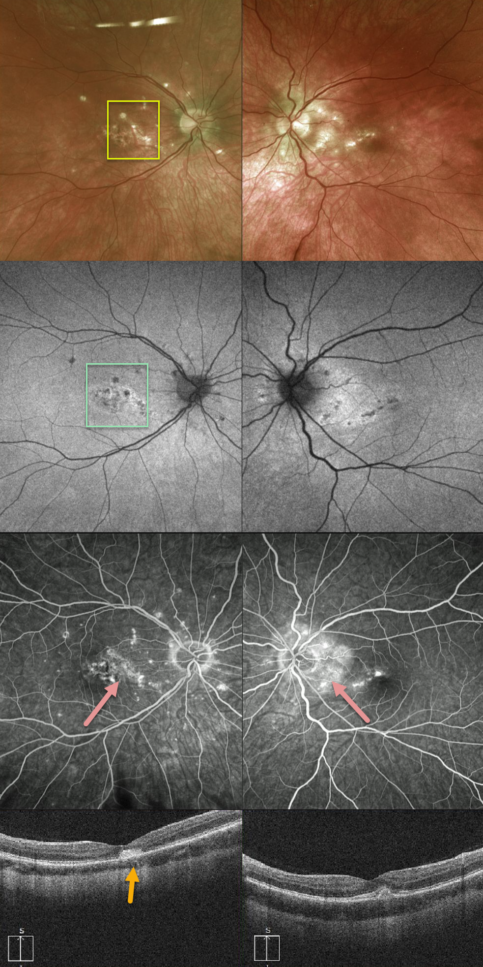
This 28-year-old patient has multifocal small bilateral macular scars with peripapillary scarring in her left eye.
The Optos photos show scattered small well-defined punctate lesions in the nasal macula with corresponding hypoautofluoresence.
Fluoroscein angiogram demonstrates early hypofluorescence of the punctate lesions with late leakage. OCT shows hump-shaped hyperreflective material with disruption of the ellipsoid zone and external limiting membrane (ELM).
Vision is counting fingers OD with inactive macular neovascularization (MNV) on chronic Eylea therapy.
Learning Points:
Punctate inner choroidopathy (PIC) is part of the multifocal choroiditis (MFC) spectrum and typically presents in young myopic females. It is never associated with vitritis.
MNV, the main cause of vision loss, can be successfully treated with anti-VEGF therapy.



