

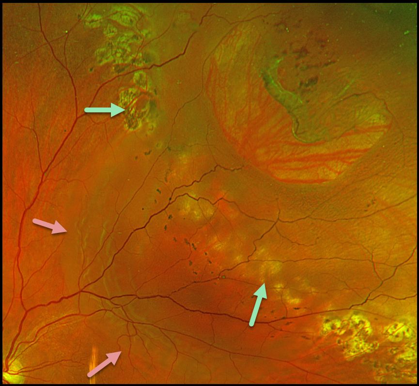
This 56YO male presented with several days of decreased vision in his left eye. He received prior treatment 1.5 years ago elsewhere for a retinal detachment. Vision was 20/30.
Optos color RG imaging shows a large lattice tear superotemporally with a surrounding rhegmatogenous retinal detachment (RRD). Several rows of appropriate and nearly confluent laser photocoagulation scarring are noted along the superonasal and inferotemporal edges of the detachment. However, barely visible depigmented laser scarring is noted more posteriorly, along with multiple tiny, 50-150 micron hyperpigmented laser scars. The detachment has broken through this region of inadequate laser.
Learning Points:
Demarcating rather than reattaching the retina is a valid option in select cases of RRD. When demarcating these detachments, it is critical that the retinopexy completely surrounds the subretinal fluid and extends to the ora; otherwise, the detachment can spread through the untreated retina, as occurred in this patient. Treatment should consist of several rows of nearly confluent, 300-500 micron photocoagulation burns. Cryotherapy or treatment with the laser indirect ophthalmoscope is often needed for the anterior treatment.
Assuming appropriate initial treatment, patients need to be followed postoperatively since the detachment can occasionally progress through the prior retinopexy. See Vrabec and Baumal, Ophthalmology 2000;107:1063-1067 for an excellent review regarding demarcation laser photocoagulation for selected macula-sparing retinal detachments.



