

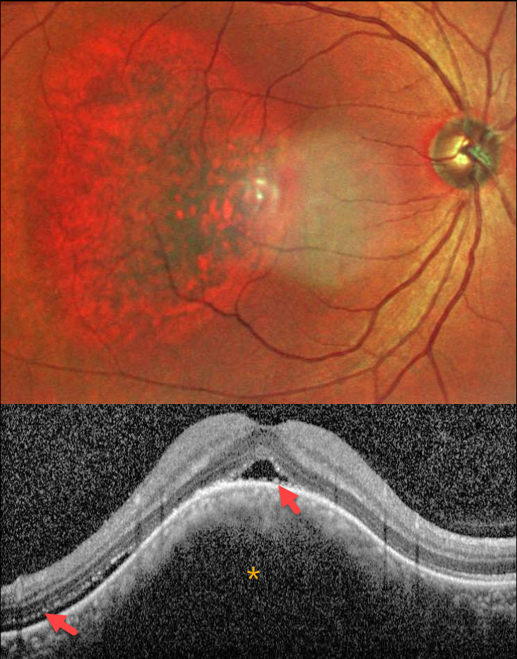
This healthy 42YO male presented with blurred and distorted vision in his right eye. Vision was 20/80 OD and 20/20 in his normal OS.
MultiColor SLO imaging shows an oval orange-red choroidal lesion in the central and temporal macula. OCT scanning shows an elevated choroidal lesion with overlying foveal and distal temporal subretinal fluid. The outer retinal layers are thinned and somewhat disorganized. Shaggy hyperreflective material overlies the central RPE.
Learning Points:
Choroidal hemangioma is a benign vascular choroidal tumor. Circumscribed tumors are isolated and non-syndromic, and appear as orange-red, dome-shaped posterior pole lesions. Diffuse hemangiomas are usually seen in association with the Sturge-Weber syndrome. The lesions are difficult to see funduscopically because the orange tumor blends with the surrounding choroid. In our patient, the MultiColor imaging more dramatically highlights this lesion.
Although there is no malignant potential, it can cause vision loss from exudation with subretinal and intraretinal fluid, which is best treated with photodynamic therapy (Yang et al, Surv Ophthalmology 2025;70:389-400).



