

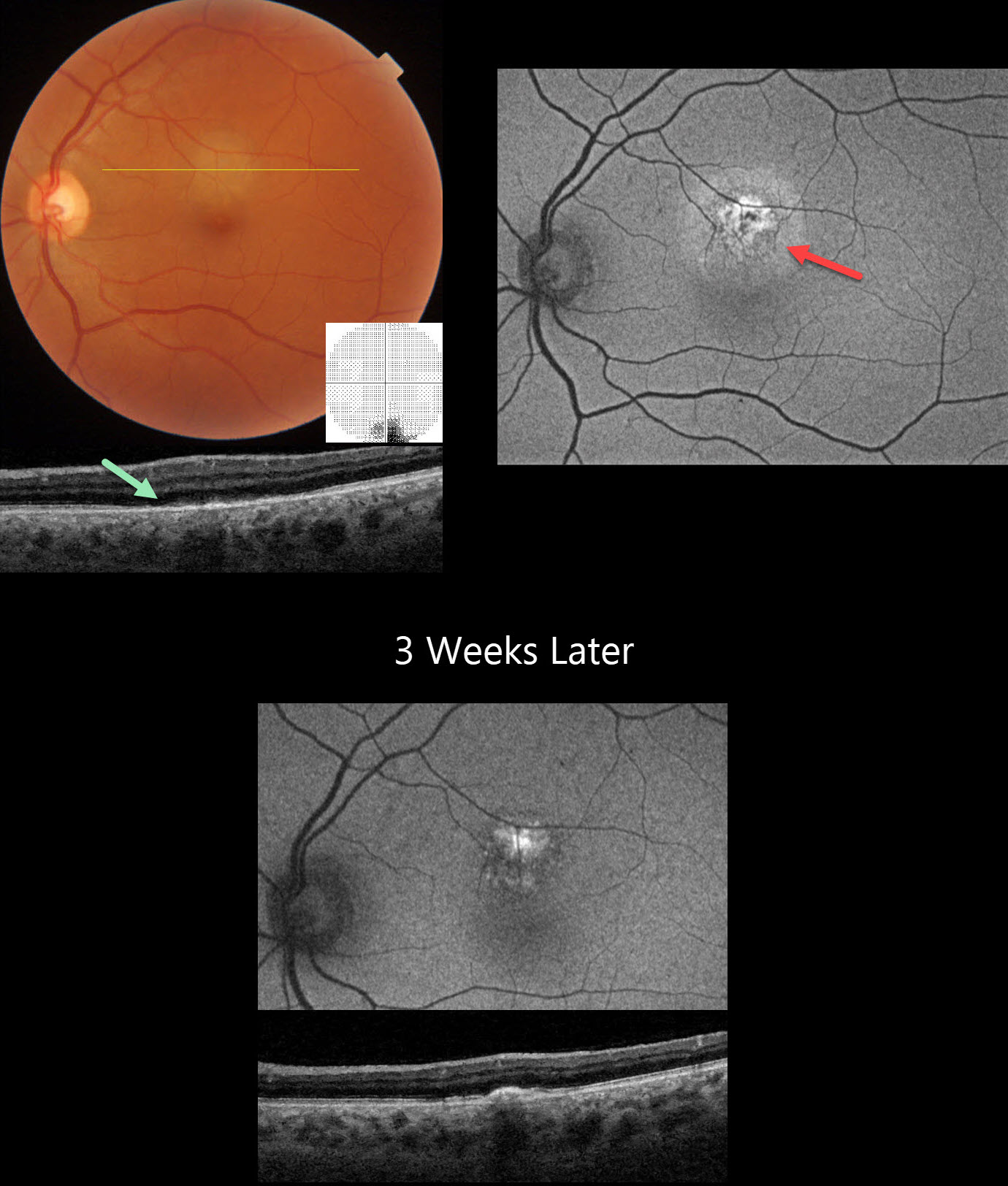
This healthy 46YO male presented with 5 days of an inferior paracentral scotoma in his left eye. Vision was 20/15 in his normal OD and 20/20 OS.
Triton color imaging shows a round, deep retinal amelanotic lesion just superior to the macular center. Swept-source OCT shows loss of the outer retinal band detail with ill-defined hyperreflective material overlying the RPE. Central 10-2 visual field shows inferior loss corresponding to the subretinal lesion. Optos fundus autofluorescence (FAF) of this lesion shows central hyper-FAF with a surrounding rim of less-intense hyper-FAF.
Coxsackie B virus antibody titers were markedly positive at >= 1:640, and observation was recommended. He returned 3 weeks later with stable symptoms and vision, although the OCT and FAF had improved.
Learning Points:
Acute idiopathic maculopathy (AIM) is an idiopathic inflammation of the outer retina, RPE, and inner choroid. There is often a preceding viral prodrome (lacking in our patient) caused by the Coxsackie virus. Healthy young patients typically present with acute unilateral moderately severe central vision loss.
The initial funduscopic changes include macular fluid and, sometimes, deep retinal blood that can mimic macular neovascularization. The OCT shows heterogeneous hyperreflective thickening of the outer retinal/RPE and fluid. Fluorescein angiography usually shows deep retinal leakage. The lesion spontaneously resolves with significant visual improvement after several weeks, usually leaving a pathognomonic bull’s-eye lesion as seen in our patient.



