

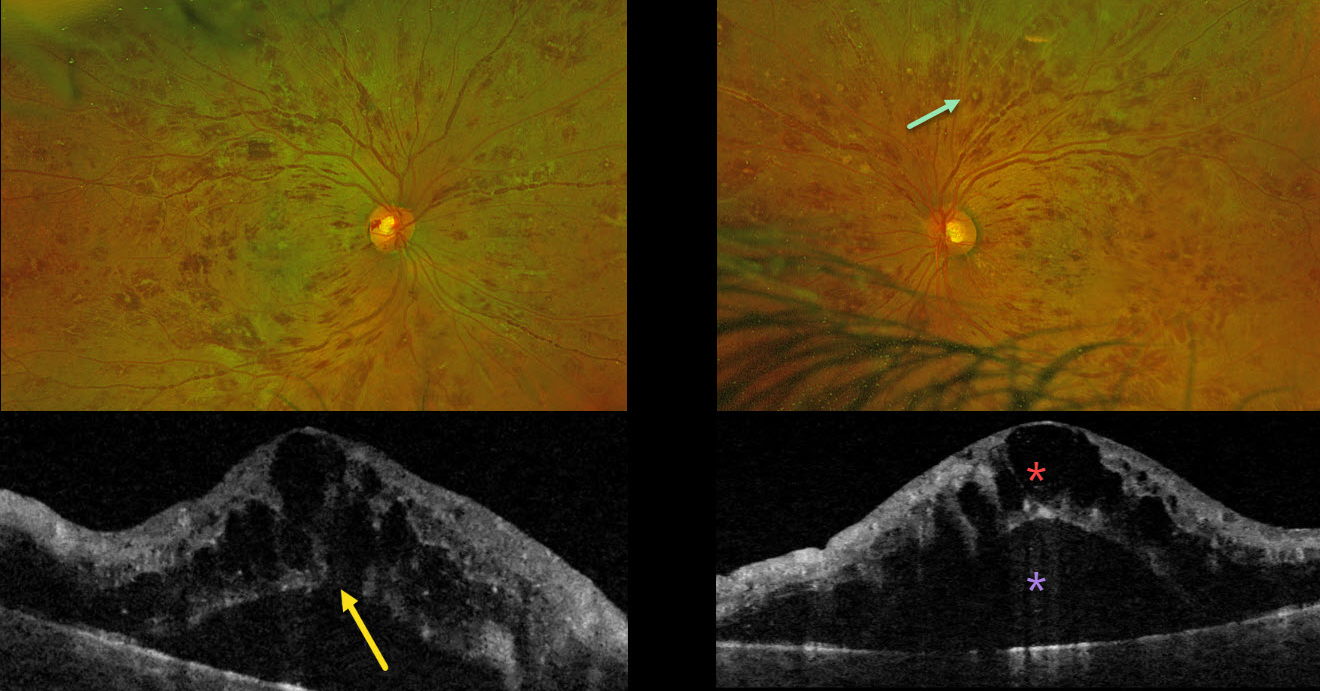
This previously healthy 38YO male presented with 1 month of bilateral blurriness. Vision was 20/60 OD and 20/80 OS.
Optos color RG imaging shows severe, bilateral, mostly white-centered retinal hemorrhages extending from the maculae into the retinal peripheries. Triton swept-source OCT shows marked bilateral cystic foveal swelling with subretinal fluid. An outer macular hole is probably present OD. His white blood cell count was markedly elevated at 206,400. Subsequent emergent oncologic workup led to the diagnosis of chronic myelogenous leukemia.
Learning Points:
The term “Roth spot” was named after Dr. Moritz Roth, who, in 1872, noted white-centered hemorrhages in patients with bacterial endocarditis. He felt that these white spots were septic emboli that originated from an infected cardiac valve. However, similar lesions can be seen in many other disorders, including leukemia, hyperviscosity, anemia, diabetes, and hypertension (Duane et al Ophthalmology 1980;87:66-69). The white material may represent a variety of causes, including fibrin. So, these lesions are best called white-centered hemorrhages, unless one is specifically referring to those found associated with bacterial endocarditis.
In our patient’s case, the severity of the retinal hemorrhages gave an appearance resembling bilateral central retinal vein occlusions (CRVO). However, CRVO hemorrhages are located predominantly in the nerve fiber layer (our patients’ hemorrhages were deep and white-centered), and the optic nerve is usually swollen with this degree of hemorrhage (our patients’ nerves were flat).



