

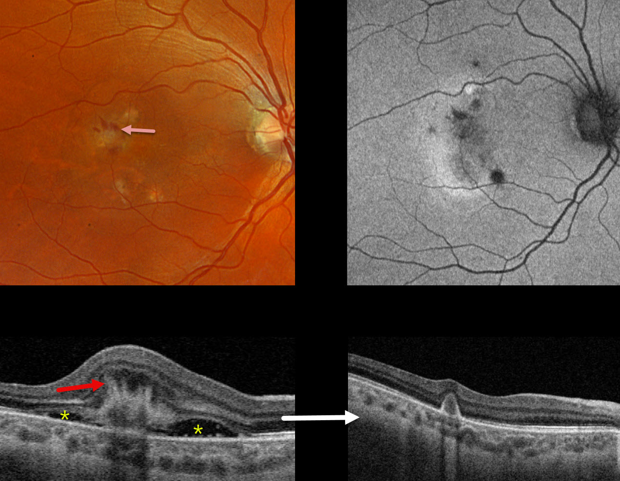
This 36YO female presented with 2 days of blurred vision in her right eye. There was no prior ocular history. Vision was counting fingers OD.
Triton color photography shows a whitish subretinal discoloration with some associated inferior subretinal fluid. A few dots of retinal blood are noted superiorly. Swept-source OCT shows a hyperreflective type 2 (above the RPE) macular neovascularization (MNV) with hyperreflective spikes radiating inwards from its inner surface. There is subretinal fluid on either edge of the MNV. Optos color RG imaging shows some small multifocal depigmented macular scars bilaterally. These scars are hypoautofluorescent.
Following an initial intravitreal Eylea HD injection, vision improved to 20/30 with complete resolution of all fluid and blood.
Learning Points:
The pitchfork sign was originally described by Hoang et al (Retina 2013;33:1049-1055) as a unique OCT finding in inflammatory MNV. Falavarajani et al more recently noted that this finding is also associated with non-inflammatory MNV (Ophthalmic Surg Lasers Imaging Retina 2019;50:719-725). The pathogenesis of the avascular pitchfork spikes is unknown but may be related to outer retinal traction or Muller cell activation.
Punctate inner choroidopathy (PIC) is part of the multifocal choroiditis (MFC) and panuveitis spectrum. MNV, the main cause of vision loss, can be successfully treated with anti-VEGF therapy, as in our patient.



