

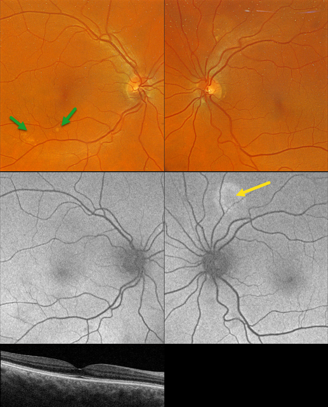
This healthy 65YO founder of Retina Rocks presented for his once-per-decade ‘eye examination’ which included an undilated slit lamp examination by his partner, Triton swept-source OCT, and Optos color RGB imaging. His mother had a history of neovascular AMD, and he has a known history of drusen. Given his family history, he has been taking AREDS2 supplementation for many years. There were no ocular symptoms, and vision was 20/20 bilaterally.
Triton color imaging shows some pachydrusen in the inferotemporal right macula. Swept-source OCT is normal except for a thickened choroid. Optos fundus autofluorescence (FAF) shows a round area of hyper-FAF superior to the left nerve.
Learning Points:
Pachydrusen were described by Spaide as a distinct pachychoroid phenotype (Retina 2018;38:708-716). Unlike typical AMD, where cone-based soft drusen are symmetrically localized to the foveal region, pachydrusen are eccentrically located, far less numerous, and have an irregular contour. Since choroidal thickness is influenced by numerous factors, including age and refractive status (Xie et al, IOVS 2022;63;2;34), these must always be considered when reviewing an individual patient’s scans. Eyes with AMD typically have age-related choroidal thinning, but eyes with pachydrusen, as in this case, are associated with a relatively thickened choroid.
Our patient became aware of his macular drusen well before pachychoroid was described by Freund in 2013 (Warrow et al, Retina 2013;33:1659-1672), and he attributed his early macular drusen to AMD. In 2025, he understands that his findings are consistent with the pachychoroid spectrum, as evidenced by the bilaterally thickened choroid, pachydrusen OD, and FAF findings suggestive of prior extramacular central serous OS. He does not have intermediate AMD, is at minimal risk for developing AMD, and thus doesn’t need AREDS2 supplementation. However, like the rest of us, he is only human and finds emotional comfort in continuing to take these supplements.



