

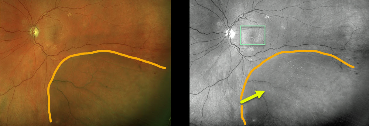
This patient with degenerative retinoschisis has extensive refractile dots, thought to be Muller cell footplates, which are well-visualized in both the Optos pseudocolor and green channel images.
This diabetic patient also has subretinal drusenoid deposits that are best seen with the green channel.
Learning Points:
Degenerative retinoschisis is virtually never prophylactically treated (see Byer 1986;93;1127-1137).
Patients can rarely develop a rhegmatogenous retinal detachment when liquid vitreous accesses the subretinal space thru both an inner and outer layer hole.



