

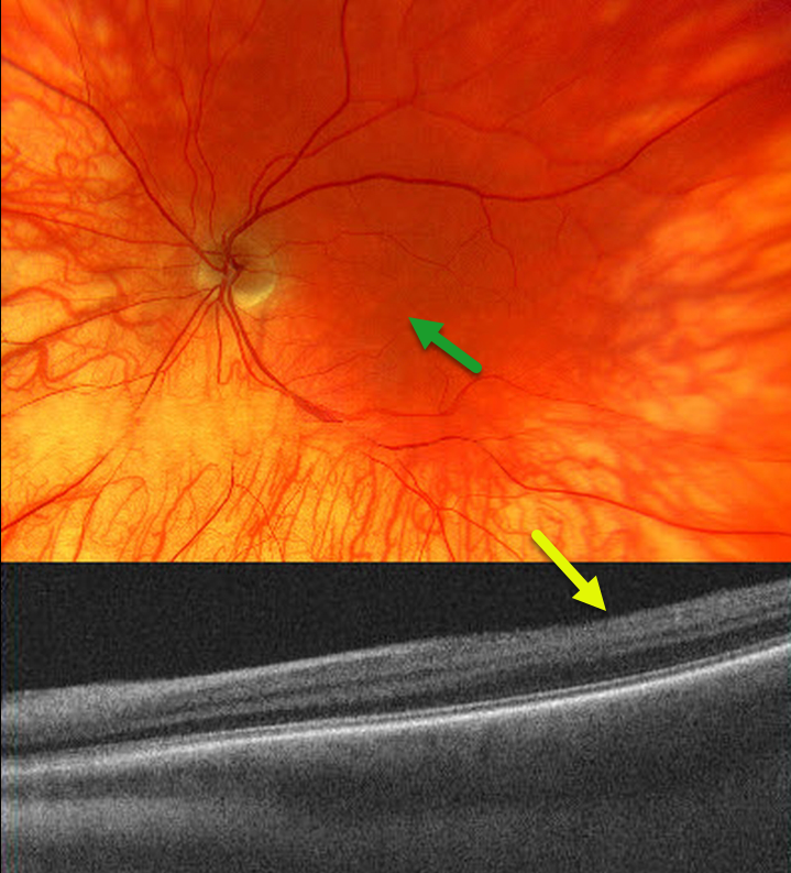
The 53YO male has a known history of ocular albinism, reporting poor visual acuity and horizontal nystagmus since childhood. Vision was 20/200 bilaterally.
Color imaging of the left eye shows a diffusely hypo-pigmented fundus with foveal hypoplasia.
OCT imaging shows a flat foveal contour (fovea plana). Similar findings were noted in the right eye (not shown).
Learning Points:
Ocular albinism is an X-linked recessive disorder that affects only the eyes and is caused by a mutation in the OA1 gene.
Findings seen in ocular albinism include strabismus, nystagmus, iris transillumination defects, blonde fundus, and foveal hypoplasia. There are often a higher number of crossed nerve fibers at the optic chiasm.
Fovea plana is characterized by the absence of the foveal pit, which is formed embryologically by the inner retinal tissues and vasculature being displaced centrifugally. It is most commonly associated with albinism, prematurity, or as an isolated finding. Despite the abnormal foveal architecture, vision is usually normal, unless due to albinism.



