

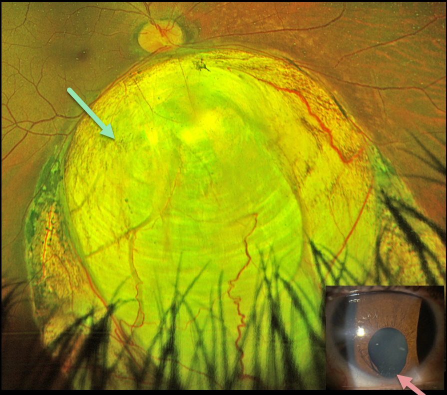
This healthy 25YO female’s primary care doctor noticed an unusual finding in her right eye and referred her to us for an examination. She said that her right pupil had looked different.
Optos color RG imaging shows a large inferior choroidal coloboma extending from the inferior disc margin into the retinal periphery. An inferior iris coloboma is noted.
Learning Points:
Choroidal colobomas are caused by failure of the optic vesicle and choroidal fissure to close during fetal development. Choroidal coloboma can present as solitary or multiple lesions, as in this case. Visual prognosis depends on the location and involvement of the optic nerve and macula.
We will follow our patient yearly due to her increased risk of retinal detachment and expect her to do well, given the normal central macular appearance.



