

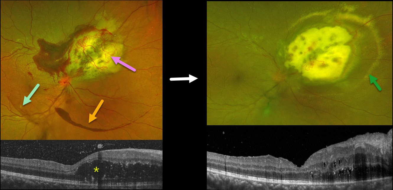
This 27YO female has been followed for an exudative astrocytic hamartoma in her right eye. Vision is 20/20 bilaterally. She was diagnosed with tuberous sclerosis at age 5 and has been followed closely by neurology.
Optos color RG imaging shows a superonasal yellow-and-white lesion with overlying neovascularization and exudates, along with areas of vitreous and subhyaloid blood.
OCT shows significant mostly outer retinal cystoid edema nasal to the fovea, as well as some vitreous blood.
She received a series of intravitreal Avastin injections with modest response and was more recently switched to Vabysmo injections. Two weeks following her third Vabysmo injection, the overlying neovascular network and blood are nearly resolved, but the surrounding subretinal lipid is increasing. The cystoid macular edema has mildly improved.
This case was submitted by Yuenpang Cheung, Stephanie Choi, and Brian Marr.
Learning Points:
Tuberous sclerosis is an autosomal dominant condition caused by mutations in the tumor suppressor genes TSC1 or TSC2. Tuberous sclerosis has multiple systemic manifestations affecting the brain (cortical tubers, subependymal nodules, epilepsy), skin (facial angiofibromas, shagreen patch, ash leaf spots), heart (cardiac rhabdomyoma), and the kidneys (angiomyolipomas).
The primary ocular manifestation is a retinal hamartoma arising from glial astrocytes. Retinal hamartomas can present as subtle gray areas in the nerve fiber layer without calcification or as prominent calcified nodules arising from the inner surface of the retina with a mulberry or fish egg-like appearance.
Although patients usually remain asymptomatic, these benign tumors can enlarge and cause macular edema, serous retinal detachment, or vitreous hemorrhage.
Exudative treatment options include anti-VEGF injections, photodynamic therapy, transpupillary thermotherapy, and radiation therapy. In cases with suboptimal response, reports of systemic mTOR inhibitors, such as Sirolimus, have shown some benefit.



