

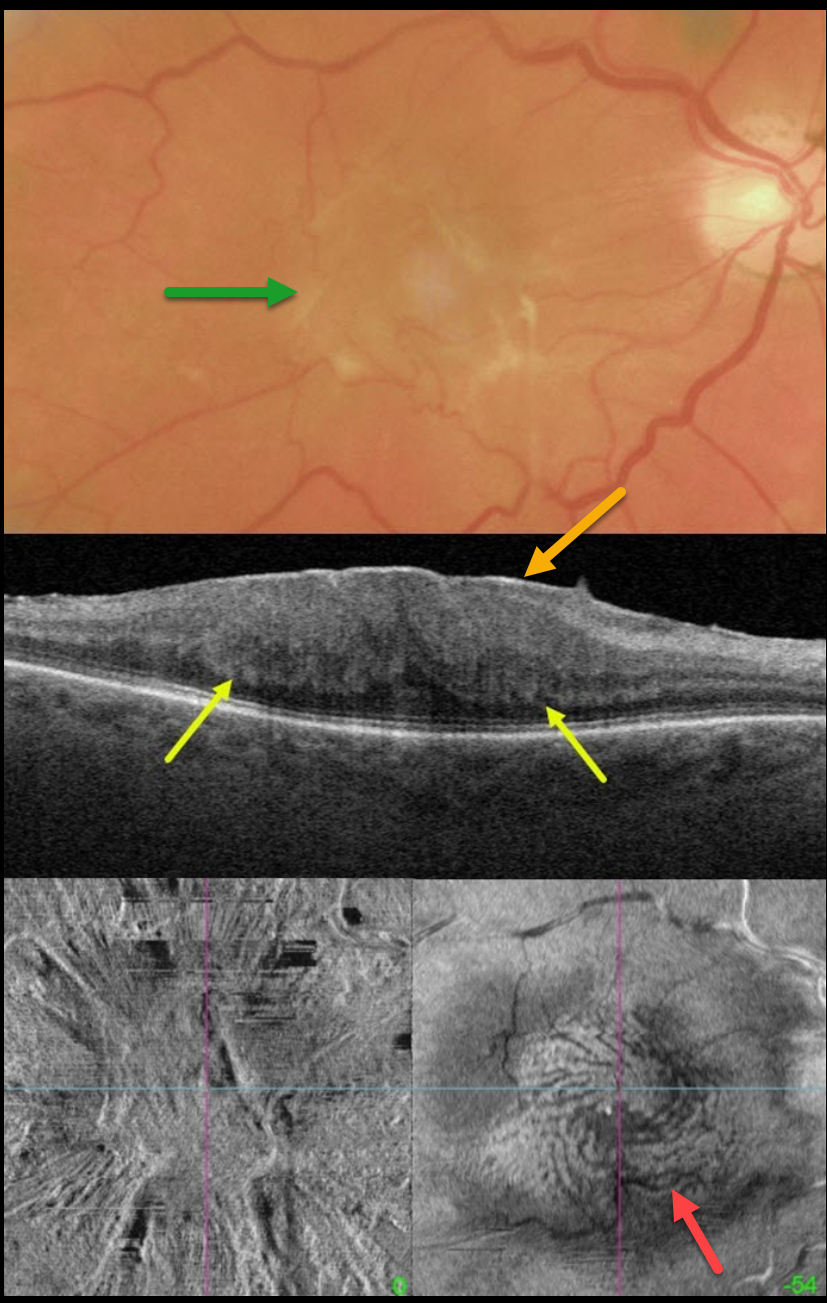
This 67YO male presented with 20/400 vision in his right eye from a symptomatic macular pucker.
Color imaging shows a severe central macular pucker. Triton swept-source OCT shows a variably adherent epiretinal membrane (ERM) with inner retinal hyperreflectivity and thickening, and complete loss of the normal inner retinal structures. The outer plexiform layer (OPL) is scalloped along its margin with the outer nuclear layer.
En face OCT of the inner retina shows variable surface distortion. En face imaging through the OPL shows a fingerprint-like whorl of hyperreflective lines.
Vitrectomy surgery was scheduled.
Learning Points:
The ‘fingerprint sign’ is a unique OCT en face finding, described by Griffin et al (Retina 2021;41:381-386), appearing as concentric, fingerprint-like waves of Henle’s layer underlying epiretinal membranes.
The authors found that the undulating OPL creates hyperreflective lines when oriented perpendicular to the incident OCT beam and hyporeflective lines when parallel to it.
In 2017, Govetto et al described an OCT finding of ectopic inner foveal layers (EIFL) associated with epiretinal membranes (AJO 2017;175:99-113). The findings range from mild underlying retinal changes (stage 1, thin ERM with foveal depression), widening of the ONL and loss of foveal depression (stage 2), and ERM continuous with EIFL crossing the entire foveal area (stage 3). All retinal layers are clearly visualized in stages 1 through 3. In stage 4, the ERM is thick, and all retinal layers are disrupted. Vision is worse with increasing stage severity. It is also associated with worse postoperative visual recovery (Yang et al, Retina 2022;42:1472-1478).



