

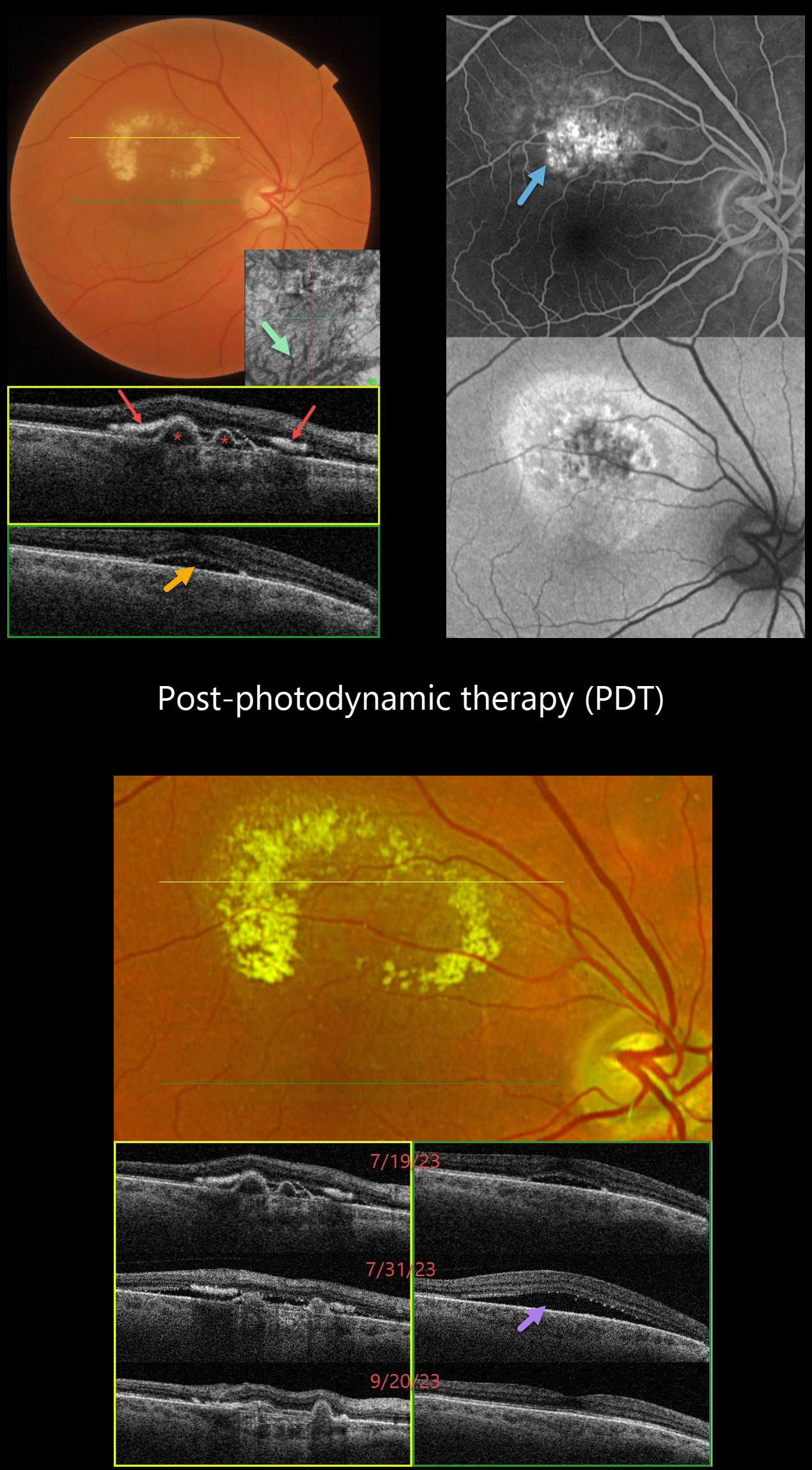
This 66YO female complained of 2 weeks of vision loss in her right eye. Vision was 20/40 OD and 20/20 in her normal OS.
Color imaging shows a circinate area of lipid in the superior macula. Triton swept-source OCT through the superior macula shows several sub-RPE polyps with hyper-reflective subretinal lipid. Scanning through the central macula shows shallow foveal subretinal fluid. Dilated inferior choroidal vessels are noted on en face OCT imaging.
Optos fluorescein angiography shows superior macular staining and leakage, with variable hyper-autofluorescence surrounding this region. Intravitreal Avastin was injected on 7/19/23, followed by photodynamic therapy (PDT) on 7/26/23.
She returned 5 days following PDT on 7/31/23 with increased blurriness. Vision remained at 20/40, although increased SRF was noted on OCT. Observation was recommended.
On 9/20/23, vision improved to 20/30, all SRF resolved, and the polyps were flattening.
Learning Points:
Polypoidal choroidal vasculopathy (PCV) is a variant of type 1 (below the RPE) macular neovascularization. It can be treated with either anti-VEGF monotherapy or combined with PDT to achieve better polyp regression and reduced treatment burden.
Transient PDT-induced increased exudative SRF sometimes develops immediately following treatment (Manayath et al, Retina 2020;40:135-144) but resolves spontaneously, as in our patient.



