

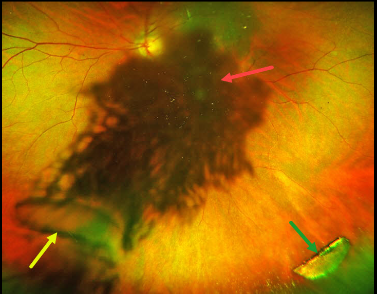
This 45YO male presented with a 4-day history of sudden vision loss in the OS immediately following penetrating trauma while working with a hammer and chisel. Vision was 20/20 in his normal OD and 20/200 OS.
Slit lamp examination OS showed a small peripheral self-sealing corneal laceration, an iris defect, and a ‘rosette cataract.’ On extreme left gaze, a semilunar defect in the posterior lens capsule and a linear defect in the anterior lens capsule were also noted.
Optos color RG imaging shows loss of central fundus detail from the cataract, the capsular defect, and a metallic intraocular foreign body (IOFB).
Fundus examination and localization of an IOFB are often difficult in the presence of secondary cataract. Ultrawide-field fundus imaging is an invaluable diagnostic tool in such cases, as it captures 200° (82% of the retina) in a single image, compared with standard fundus photography.
The rosette cataract in our patient is caused by seepage of aqueous humor through the broken lens capsule and between the lens fibers, resulting in opacification.
Urgent pars plana lensectomy, vitrectomy, removal of the IOFB, and secondary intraocular lens were performed. Vision was 20/40 six weeks following surgery.



