

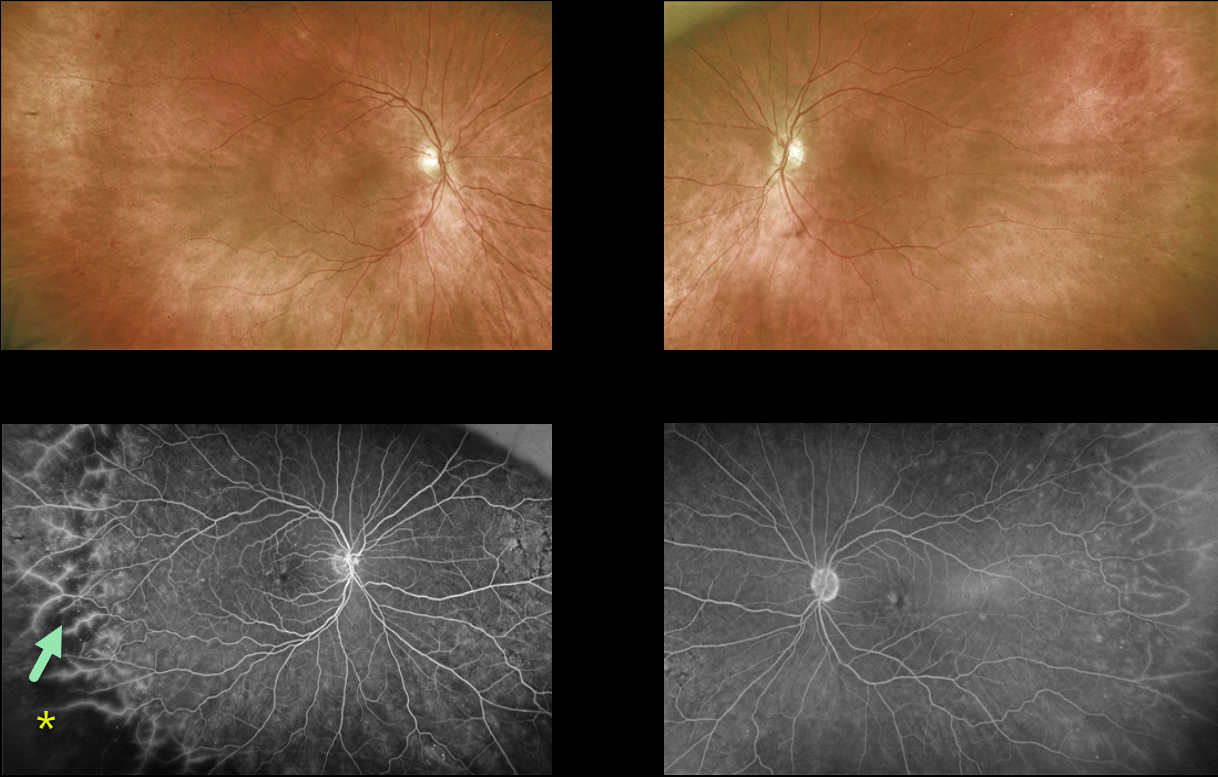
This 69YO female with a history of type 2 diabetes was referred for asymptomatic retinal changes. Vision was 20/25 OU.
Optos color RG imaging shows virtually normal posterior poles with temporal peripheral retinal hemorrhages.
Fluorescein angiography shows temporal ischemia with diffuse vascular staining.
Learning Points:
The DRCR Retina Network’s Protocol AA found that ultrawidefield (UWF) imaging, compared to the standard ETDRS 7-field images, increases the frequency of diabetic retinopathy identification by nearly two fold (JAMA Ophthalmology 2019;137:65-73).
UWF fluorescein angiography further increases the sensitivity for assessing retinal ischemia, with about 70% of nonperfusion located outside the posterior pole (Retina 2022;42:1302-1310).
Finally, eyes with angiographically predominantly peripheral lesions, as in our patient, are at a higher 4-year risk of worsening disease, independent of the baseline diabetic retinopathy severity score (JAMA Ophthalmology 2022;140:946-954).
UWF fundus imaging has transformed the field of retina. In our practice, we are constantly humbled by the superiority of UWF imaging in finding peripheral pathology compared to our clinical examination. And increasingly, this technology is becoming the gold standard for identifying pathology and guiding our treatment decisions.



