

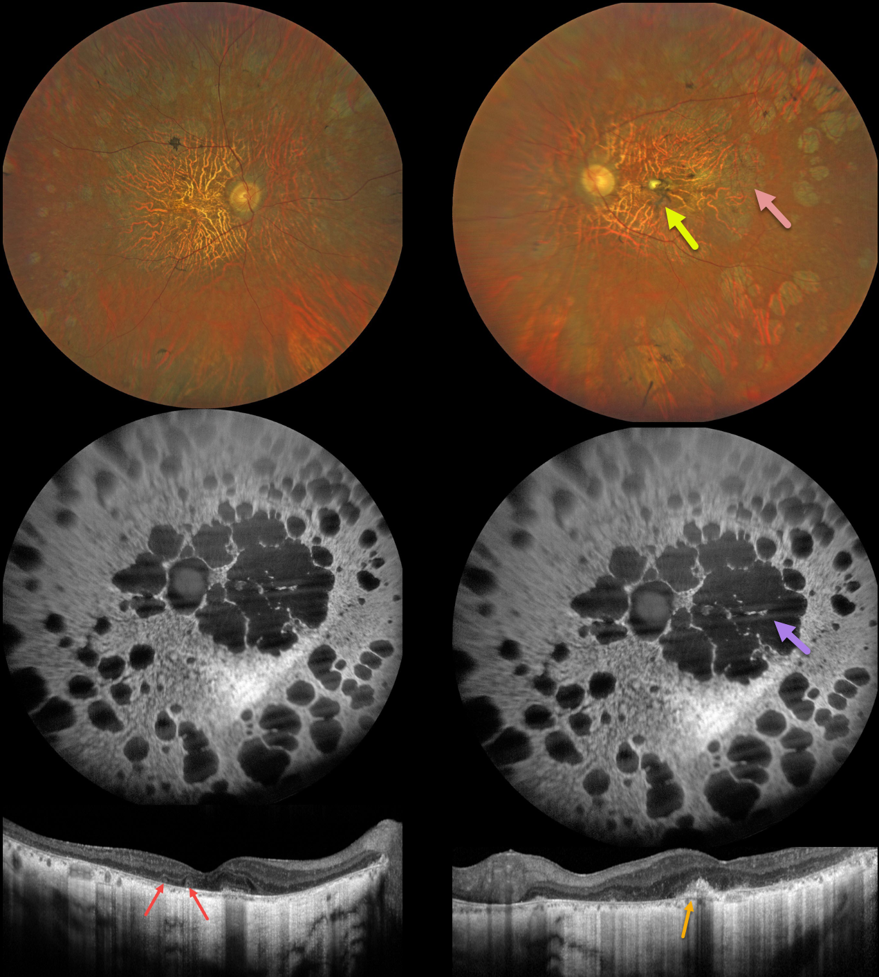
This 56YO male has a history of nyctalopia since childhood with a known mutation in the RDS/peripherin gene. Vision was 20/60 in the right eye and counting fingers in the left eye. The patient has been taking vitamin A supplementation. He has an older brother with a more advanced inherited retinal disorder (IRD).
Fundus imaging shows bilateral diffuse areas of retinal pigment epithelial atrophy. In addition, there is a yellow elevated dome-like lesion with surrounding pigment in the left fovea.
Fundus autofluorescence (FAF) shows hypo-FAF in the areas of atrophy. OCT shows diffuse outer retinal/RPE atrophy with choroidal thinning. In addition, there are a few outer retinal tubulations OD and a subretinal hyperreflective elevated lesion OS. Electrophysiology revealed extinguished rod and diminished cone responses.
Learning Points:
Mutations in the RDS/peripherin gene have been implicated in a number of inherited conditions, including retinitis pigmentosa, pattern dystrophy, retinitis punctata albescens, cone-rod dystrophy, and adult-onset vitelliform macular dystrophy.



