

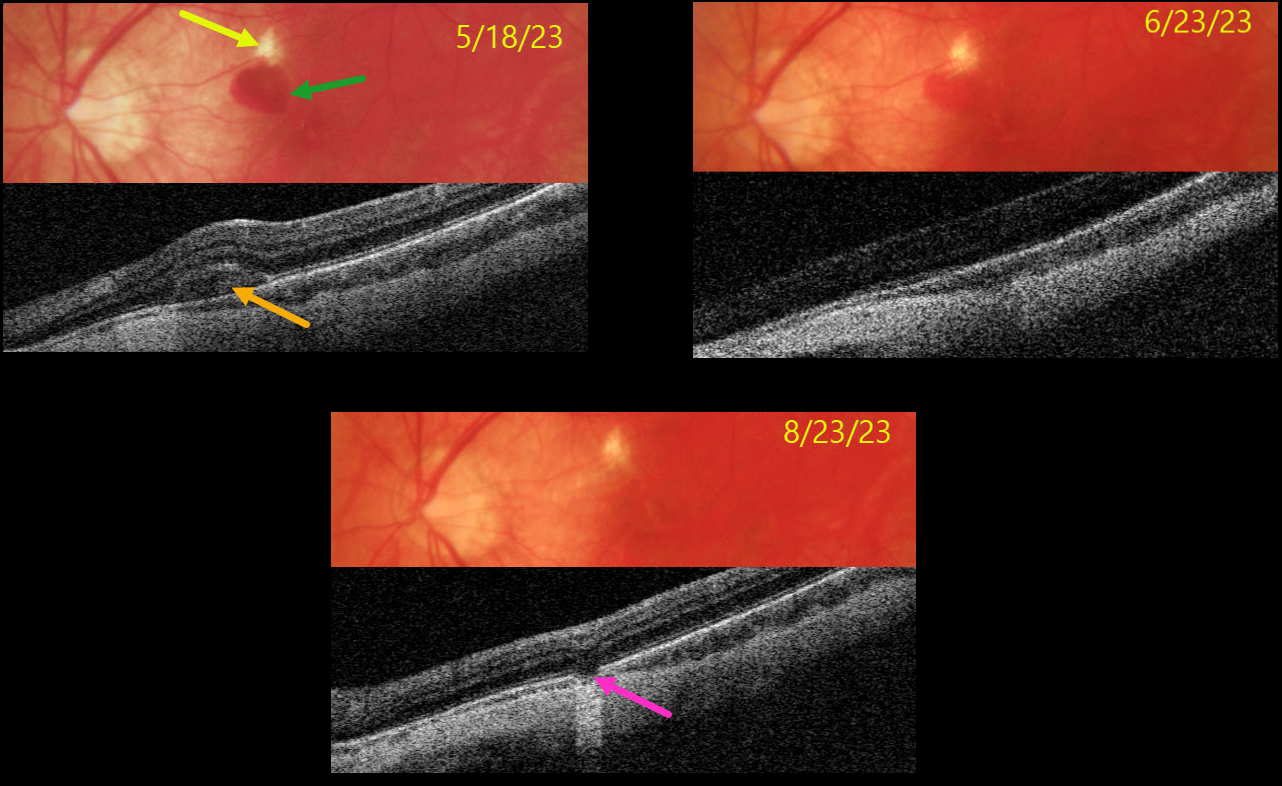
This 33YO highly myopic male presented on 5/18/23 with vision loss in his left eye. Vision was 20/40.
Color photography shows a superior macular lacquer crack just superior to a round subretinal hemorrhage. The blood is hyperreflective on Triton swept-source OCT. The blood gradually absorbed with observation.
When last examined on 8/23/23, the blood had completely resolved, and vision remained at 20/40, but with resolved symptoms. OCT scanning through the lacquer crack shows a defect in the outer retinal bands and RPE.
Learning Points:
Lacquer cracks are a common finding in patients with high myopia. They consist of breaks in Bruch’s membrane due to the outpouching/stretching of the pathologically myopic eye and increase the risk for macular neovascularization. In some cases, similar to this patient, self-limiting small subretinal avascular hemorrhages occur that do not require anti-VEGF therapy.



